


Frontal bone
The frontal bone , os frontale, in an adult forms the anterior part of the cranial vault and partly its base. It consists of four parts: frontal scales, two orbital parts and a bow.

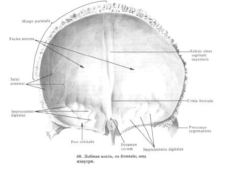
The frontal scales, squama frontalis, convex anteriorly, have the following surfaces: external, or frontal, two temporal, or lateral, and internal, or cerebral.
Outer surface, facies externa, smooth, convex anterior. On the middle line, elevation is not always noticeable - the metopic suture, sutura metopica, is the trace of the fusion of the half of the frontal bone in the early childhood. In the anterior regions, the frontal surface of the scales passes into the orbital surface, facies orbitalis, forming on each side the supraorbital margin, margo supraorbitalis, which is the upper part of the orbital margin, margo orbitalis. Above and parallel to the supraorbital margin, an arcuate elevation appears more or less prominently: the superciliary arc, arcus superciliaris. Above each brow, a round elevation is visible - the frontal tuber, tuber frontale. Between the protuberances of the superciliary arches and slightly higher, their surface of the frontal scales in the region of the nadperium has the appearance of a somewhat deeper area - glabella , glabella. The inner third of the supraorbital margin has a small supraorbital incision, incisura supraorbitalis. This tenderloin is highly variable and can be expressed in the form of a supraorbital foramen, foramen supraorbitale. Nearer to the median line, i.e. medial, lies an equally prominent frontal incision, incisura frontalis (in the supraorbital incision there pass the lateral branch of the supraorbital nerve and the vessels, in the frontal - the medial branch of the same nerve and the vessels). In the place of the above notch, a frontal foramen, foramen frontale, can form.
The lateral supraorbital margin passes into a blunt, triangular-shaped zygomatic process, processus zygomaticus; Its jagged edge connects with the frontal process of the malar bone, forming the frontal-zygomatic suture, sutura frontozygomatica.
From the zygomatic process up and back, the temporal line is directed in an arch, linea temporalis; It separates the frontal surface of the scales from the temporal surface. The temporal surface, facies temporalis, is the anterior margin of the temporal fossa, fossa temporalis, where the front tufts of the temporalis begin.
Inner surface, facies interna, concave. It has weakly expressed finger-like impressions, impressiones digitatae, and unstable arterial furrows, sulci arteriosi (as an imprint of the relief of the adjacent brain and vessels ).
In the middle of the inner surface of the frontal scales passes the furrow of the superior sagittal sinus, sulcus sinus sagittalis superioris. Its both edges, going up and back, pass into the eponymous furrow of the parietal bones, and below connect in the acute frontal crest, crista frontalis (to it is attached the dura of the dura mater - the sickle of the big brain). The lowest part of the ridge and the wing of the crock of the trellis bone, ala cristae galli ossis ethmoidalis, form a canal - a blind hole, foramen cecum, in which a vein draining blood from the nasal cavity to the superior sagittal sinus.
Upper or posterior margin of frontal scales - parietal margin, margo parietalis, thickened; Its jagged edge connects with the frontal edge of the parietal bones, forming a coronal suture, sutura coronalis. The lower portions of the triangle-shaped scales are connected to the frontal margin of the large wings of the sphenoid bone .
Each orbital part, pars orbitalis, of the frontal bone is part of the upper wall of the orbit. From the supraorbital margin of the frontal scales, it is directed backwards and horizontally. It distinguishes the lower ophthalmic and upper cerebral surfaces.
Glazed surface, facies orbitalis. Inverted into the orbit cavity, smooth and concave. In the lateral part of her, at the base of the zygomatic process, lies a shallow fossa of the lacrimal gland, fossa glandulae lacrimalis, the location of the lacrimal gland.
In the medial part of the orbital surface there is a weakly expressed block fovea, fovea trochlearis, near which there is often a cartilaginous block awn, spina trochlearis (here a cartilaginous ring is attached, which is a block of the tendon of the upper oblique muscle of the eyeball). The upper cerebral surface, facies cerebratis, of the ophthalmic part has well-defined imprints of the adjacent surface of the frontal lobes of the brain in the form of finger impressions, impressiones digitatae (gyrorum).

The glabellar parts divide from each other the trellis, incisura ethmoidalis, in which the trellis plate, lamina cribrosa, of the latticed bone is located. The cutting along the sides is limited by the edge, outside of which there are dimples that cover the open cells of the upper part of the maze of the latticed bone, forming their upper wall. Between the latticed dimples pass two lateral grooves in the transverse direction - anterior and posterior, which together with the same grooves of the labyrinth of the lattice bone form tubules. The latter open on the inner wall of the orbit - that two small holes: a foramen ethmoidale anterius (through which the front latticed vessels and nerve pass), and a posterior latticed aperture, foramen ethmoidale posterius (through which the posterior latticed vessels and nerve pass). The edge of the trellis is connected with the upper edge of the orbital plate, lamina orbitalis, of the trellis, forming the frontotal suture, sutura frontoethmoidalis, and in front - with the tear bone - the frontal lacrimal suture, sutura frontolacrimalis.
The posterior edge of the orbital part, which is rakish and serrate, connects with the small wing of the sphenoid bone, forming the inner portion of the wedge-frontal suture, sutura sphenofrontalis.
The lateral edge of the orbital part is scabrous, triangular in shape. It joins the frontal margin of the large wing of the sphenoid bone and forms the outer portion of the wedge-frontal suture.
The nasal part, pars nasalis, of the frontal bone in the form of an arc closes the front of the trellis. Ahead, in the middle of the nose, a forward (sometimes double) obliquely downward and forward nasal spine, spina nasalis, pointed at the tip and flattened laterally. It is surrounded at the front and side by a serrated nasal margin, margo nasalis. It connects with the upper edge of the nasal bone, forming a frontal-nasal suture, sutura frontonasalis, and with the frontal processus frontalis, the maxilla, forming the frontal-maxillary suture. Sutura frontomaxillaris. The lower surface of the posterior sections of the nose has shallow dimples, which, as noted, cover the open cells of the maze of labyrinths of the latticed bone.
On each side of the nasal awning there is one aperture of the frontal sinus, apertura sinus frontalis; Going up and anterior, it leads into the cavity of the corresponding frontal sinus. The frontal sinus, sinus frontalis, is a pair of cavities lying between the two plates of the frontal bone in its anterior regions. The frontal sinus
They take it in the airy bones of the sinuses. The right sinus from the left separates the vertical septum of the frontal sinuses, septum sinuum frontalium, Deviating to the side, the septum causes an unequal size of the cavities of both sinuses. The boundaries vary dramatically. Sometimes the frontal sinuses reach up to the frontal tubercles, down to the supraorbital margin, posterior to the small wings of the sphenoid bone and to the side to the cheekbones. The aperture of the frontal sinus connects the frontal sinus and the middle nasal passage, meatus nasi medius, of the nasal cavity. The sinus cavity is lined with a mucous membrane.
You will be interested to read this:

Comments
When commenting on, remember that the content and tone of your message can hurt the feelings of real people, show respect and tolerance to your interlocutors even if you do not share their opinion, your behavior in the conditions of freedom of expression and anonymity provided by the Internet, changes Not only virtual, but also the real world. All comments are hidden from the index, spam is controlled.