


Pleura. Structure, functions
Pleura.
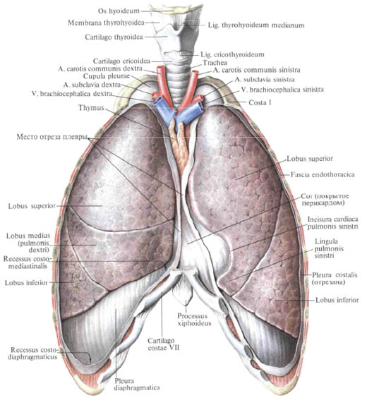
The lungs are covered with pleura, pleura. It, like the peritoneum, is a smooth, shiny serosa, tunica serosa. There are parietal pleura, pleura parietalis, and visceral (pulmonary), pleura visceralis (pulmonalis), between which a gap is formed - the pleural cavity, cavitas pleuralis, filled with a small amount of pleural fluid.
The visceral (pulmonary) pleura directly covers the parenchyma of the lung and, being tightly intertwined with it, extends into the depths of the interlobar furrows.

The parietal pleura is fused to the walls of the thoracic cavity and forms the rib pleura, pleura costalis, and the diaphragmatic pleura, pleura diaphragmatica, and the mediastinal pleura mediastinalis, which bounds the mediastinal walls. In the area of the lung gates, the parietal pleura turns into pulmonary, covering the transitional fold with the root of the lung in front and behind.
Below the lung root, the transitional fold pleura forms a duplicate - a pulmonary ligament, lig. Pulmonale.
In the region of the apex of the lungs, the parietal pleura forms the dome of the pleura,
Cupula pleurae, which in the upper parts is dorsally attached to the head of the 1st rib, and anterolateral surface adjoins the stair muscles.
Parts of the pleural cavity in the form of an acute angle between two parietal sheets, passing from one wall to the other, are called pleural sinuses, recessus pleurales.
Distinguish the following sines:
1) rib-diaphragmatic sinus, recessus costodiaphragmaticus, located at the site of the transition of the costal pleura into the diaphragmatic;
2) costal mediastinal sinuses, recessus costomediastinales, are formed at the sites of the transition of the rib pleura to the mediastinal pleura; The anterior sinus behind the sternum, the posterior sinus, less pronounced, in front of the vertebral column;
3) diaphragmmediastinal sinus, recessus phrenicomediastinalis, lies in the place of the transition of the mediastinal pleura into the diaphragmatic one.
The lower border of the lungs does not coincide with the boundaries of the parietal pleura.
The lower border of the parietal pleura passes: along the linea mediana anterior - on the VI-VII rib; By linea medioclavicularis (mamillaris) - on the 7th rib (lower edge); By linea axillaris media - on X edge; By linea scapularis - on the XI-XII edge: linea paravertebralis - on the XII edge.
Thus, the depth of the rib-diaphragmatic sinus is greatest in linea axillaris media.
The anterior border of the parietal pleura of both lungs extends from the sternoclavicular joints down behind the handle and the body of the sternum to the lower edge of the sternal ends of the IV ribs. Here, the anterior edge of the pleura of the right lung continues down to the intersection of the VI rib with the linea mediana anterior, and the left lung at the level of the IV rib turns to the left and, describing the arc of the intersection of the VII rib with linea medioclavicularis.
On their way, the front edges of the parietal pleura of both lungs diverge in the upper and lower divisions and form a triangular space behind the sternum, in which the thymus gland is located, and in the lower part a triangular slit, bounded posteriorly by the pericardium.
You will be interested to read this:

Comments
When commenting on, remember that the content and tone of your message can hurt the feelings of real people, show respect and tolerance to your interlocutors even if you do not share their opinion, your behavior in the conditions of freedom of expression and anonymity provided by the Internet, changes Not only virtual, but also the real world. All comments are hidden from the index, spam is controlled.