


A throat of the person. Function, pharynx structure
Pharynx , pharynx, is part of the digestive tube through which the food lump from the oral cavity moves into the esophagus. At the same time, the pharynx is the path through which air passes from the nasal cavity to the larynx and back.
The pharynx is located in front of the cervical vertebral column , its posterior wall adjoins the pre-vertebral plate of the cervical fascia and extends from the base of the skull to the sixth cervical vertebra , where it tapers to the esophagus. The length of the pharynx is 12-15 cm.
The upper part of the pharynx communicates with the nasal cavity and is called the nasal part of the pharynx (nasopharynx), pars nasalis pharyngis. It corresponds to the 1st and 2nd cervical vertebrae . Its middle part communicates with the oral cavity and is called the oral part of the pharynx, pars oralis pharyngis. The posterior wall of this part of the pharynx corresponds to the body of the third cervical vertebra. The lower part is located behind the larynx - this is the throat part of the pharynx, pars laryngea pharyngis. Its posterior wall corresponds to the level of IV-VI cervical vertebra.
The walls of the pharynx.
In the throat distinguish the upper wall - the arch of the pharynx, the fornix pharyngis, the anterior, posterior and two lateral walls. The pharynx is attached to the outer surface of the base of the skull along a line running from the pharyngeal tubercle to the outer orifices of the carotid canals and further forward to the base of the medial plates of the pterygoid processes of the sphenoid bone . In this part of the throat is inactive, because it is fused with the bony formations of the skull. In the lower part of the pharynx, due to the surrounding well-developed, loose connective tissue, it is very mobile.
The pharyngeal walls are formed by the mucosa, the submucosa, the muscular membrane and adventitia.
The mucous membrane, tunica mucosa, is covered in the nasopharynx by ciliated epithelium, and in the middle and lower regions it is multilayered. It is an extension of the mucous membrane of the nasal cavity and mouth, and below it passes into the mucous membrane of the larynx and esophagus.
The submissive base, tella submucosa, is represented by a dense connective tissue plate, which is denser in the upper parts of the pharynx and is called the pharyngeal basilar fascia, fascia pharyngobasilaris. Densely fused with it, the mucous membrane of the pharynx does not form folds. Only in the lower parts of the pharynx the submucosa is constructed from loose connective tissue, so that the mucous membrane of the pharynx forms a series of longitudinal folds. In the submucosa base there are various sizes and forms of pharyngeal glands, glandulae pharyngeae, the ducts of which open on the surface of the mucous membrane. In various parts of the submucosa of the pharynx, considerable accumulations of lymphoid tissue form.
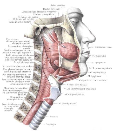
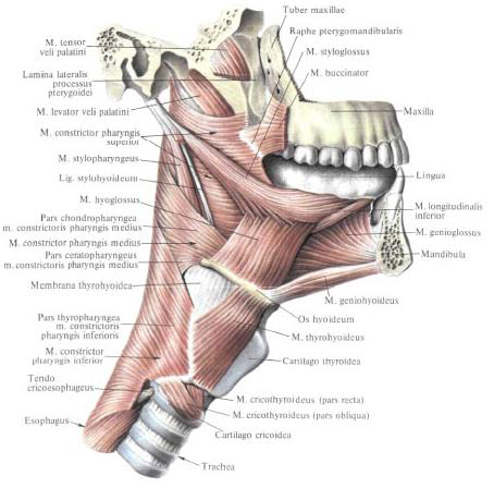
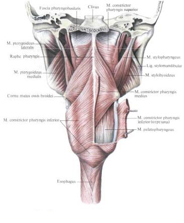
The muscular membrane of the pharynx, tunica muscularis pharyngis, is formed by five pairs of striated muscles. Three of these are muscles that constrict the pharynx (constrictors), mm. Constrictores pharyngis, going in the transverse direction. All three pairs of these muscles meet posteriorly along the median line, partially passing to the opposite side and weaving into the longitudinally located connector of the weaved bundle, starting from the pharyngeal tubercle and bearing the name of the pharyngeal suture, the raphe pharyngis. The remaining two pairs of muscles are the muscles that lift the pharynx. They go in the longitudinal direction.
The muscles of the pharynx include:
- The upper pharyngeal constrictor, m.constrictor pharyngis superior, has the shape of a quadrangular plate. It starts from several sections, so that it is divided into four parts:
- The pharyngeal part, pars pterygopharyngea, begins from the hook and posterior edge of the medial plate of the pterygoid process;
- The bucco-pharyngeal part, pars buccopharyngea, begins from the wing-jaw joint, raphe pterygomandibutaris. This part of the pharynx is covered by the buccal-pharyngeal fascia, fascia buccopharyngealis, passing from the buccal muscle;
- The maxillo-pharyngeal part, pars mylopharyngea, starts from the posterior end of the maxillo-hyoidal line of the body of the lower jaw;
- The glossopharyngeal part, pars glossopharyngea, begins from the root of the tongue .
Muscle bundles go horizontally along the side wall of the pharynx to the posterior and connect to the bundles of the muscle of the opposite side in the throat of the pharynx. The upper edge of the muscle does not reach the base of the skull , and the site of the pharyngeal wall, devoid of the muscle layer, consists of a thickened fibrous base of the pharynx - a pharyngeal basilar fascia, fascia pharyngobasilaris.
- Middle pharynx constrictor, m. Constrictor pharyngis medius, consists of two parts:
- The cartilaginous part, pars chondropharyngea, originates from the small horn of the hyoid bone ;
- The coronary-pharyngeal part, pars seratopharyngea, starts from the large horn of the hyoid bone.
The muscle has the form of a triangular plate, the broad base of which is located along the seam of the pharynx, and the apex is facing the hyoid bone . The upper tufts partially cover it. Constrictor pharyngis superior.
- Lower pharynx constrictor, m. Constrictor pharyngis inferior, flat, covers partially m. Constrictor pharyngis medius, consists of two parts:
- The pharyngopharyngeal part, pars thyropharyngea, begins from the outer surface of the plate of the thyroid cartilage of the larynx;
- The ring-pharyngeal part, pars cricopharyngea, starts from the lateral surface of the cricoid cartilage of the larynx.
Muscular bundles, diverging in a fanlike manner, are joined together with the suture of the pharynx with the bundles of the same muscle of the opposite side.
Function: all muscles narrow the lumen of the pharynx.
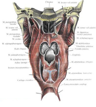
- Shillopharyngeal muscle, m. Stylopharyngeus, narrow, long, starts from the styloid process of the temporal bone, goes down along the pharyngeal wall, penetrates it between m. Constrictor pharyngis superior and m. Constrictor pharyngis medius, and, dividing into bundles, weaves into the pharyngeal wall, and some of the bundles reach the cartilage of the larynx.
Function: raises pharynx and larynx.
- The neo-pharyngeal muscle, m. Palatopharyngeus.
From the outside, the muscular membrane of the pharynx is covered by the connective tissue membrane - adventitia, adventitia that passes into the connective tissue (adventitial) envelope of the esophagus.
The cavity of the pharynx.
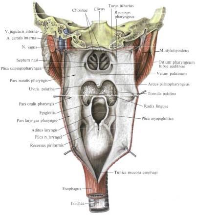
The nasal part of the pharynx, pars nasalis pharyngis, extends from the pharyngeal arch to the soft palate . In front of her open the back holes of the nasal cavity - choana, choanae.
On the side wall on each side, at the level of attachment of the posterior end of the inferior nasal shell, is a funnel-shaped pharyngeal opening of the auditory tube, ostium pharyngeum tubae auditivae. Through the auditory tube, the pharyngeal cavity is connected to the middle ear cavity.
In the region of the pharyngeal opening of the auditory tube, the mucosa forms two folds above the opening. In the back fold, the cartilage of the auditory tube is formed, forming a tube roller, torus tubarius. The tube roller continues into a gradually thin fold of the mucous membrane - a tubal-pharyngeal fold. Plica salpingopharyngea. A cushion from the pharyngeal opening of the auditory tube is determined by the muscle cushion that lifts the palatine curtain, torus levatorius. From the anterior end of the tube roll to the soft palate stretches the palatal-palatal fold, plica salpingopalatina. Behind him there is a small depression - pharyngeal pocket, recessus pharyngeus.
Lymphoid tissue of the nasal part of the pharynx forms aggregations - pharyngeal (adenoid) tonsil, tonsilla pharyngealis (adenoidea), and two tubal tonsils, tonsilae lubariae. The pharyngeal tonsil has the same structure as the palatine tonsil. Its surface, turned into the pharynx cavity, is covered with amygdala dimples, fossulae tonsillares, into which open sinuous indentations - amygdala crypts, cryptae tonsillares, located in the stroma of the amygdala. In the middle of the medial pocket of the pharyngeal tonsil, the diverticulum - pharyngeal bag, bursa pharyngealis - is sometimes opened. Pipe tonsil is a group of follicles running around the pharyngeal opening of the auditory tube.
Tonsils: lingual, palatine, tubal and pharyngeal - together form the lymphoepithelial ring .
The oral part of the pharynx, pars oralis pharyngis, extends from the level of the soft palate to the entrance to the larynx. When swallowing, the cavity of the oral part of the pharynx is separated from the cavity of its nose by a soft sky, assuming a horizontal position. In front, the oral cavity of the pharynx through the throat isthmus communicates with the oral cavity,
The throat part of the pharynx, pars laryngea pharyngis, extends from the entrance to the larynx to the lower edge of the cricoid cartilage of the larynx, at the level of which it passes into the esophagus. On the front wall is the entrance to the larynx, aditus laryngis, through which the cavity communicates with the cavity of the larynx, cavitas laryngis. In the laryngeal part of the pharynx, between the inner surface of the thyroid cartilage of the larynx and the scapular-fold fold, there is a depression - plica aryepiglottica, - a pear-shaped pocket, a recessus piriformis. Here, the mucosa forms a fold of the laryngeal nerve, plica nervi laryngei, above the upper laryngeal nerve, laryngeus superior.
Ocroglotocious fiber.
The pharynx is surrounded by a considerable amount of loose connective tissue filling the peripheral cellular space, spatium peripharyngeum. It is divided into the zygopharyngeal and lateral okolohlotchnye spaces.
The pharyngeal space, spatium retropharyngeum, is a gap bounded from the front by the buccal pharyngeal fascia, fascia buccopharyngealis, and posteriorly by a prevertebral plate of the cervical fascia, lamina prevertebratis fasciae cervicalis. Above, the swallowing space reaches the base of the skull, and downwards passes into the posterior-vascular space, the spatium retroviscerale, the neck. Oropharyngeal lymph nodes are located in the pharynx.
Lateral peripheral space, spatium lateropharyngeum, paired. The side wall of the pharynx and the muscle that lifts the palatine curtain make up its medial wall. The lateral wall of the space is formed by a muscle that strains the palatine curtain, the medial pterygoid muscle and fascia, stretched between the posterolar edge of the medial pterygoid muscle, the base of the skull and the subulate spine. From the front, both walls converge and the cell space is closed by the transition of the fascial plate from the buccopharyngeal fascia to the medial pterygoid muscle. Between the muscle that lifts and the muscle that strains the palatal curtain, the fiber of the lateral okolohlotoknogo space is communicated with the accumulation of fiber under the mucous membrane Amygdala fossa, labial-lingual and pterygo-mandibular folds. At the top, the lateral occipital space reaches the base of the skull, and from the bottom is limited by the fascial case of the submaxillary gland.
A dense fascia, stretched between the styloid process, the base of the skull, the shillopharyngeal muscle and the pharyngeal wall, serves as a boundary between the retropharyngeal and lateral peripheral spaces. This fascia envelops the internal carotid artery , the internal jugular vein and the vagus nerve, continuing on the neck in the form of a sleepy vagina, vagina carotica
Innervation: plexus phyrangeus formed by n. Glossopharyngeus, n. Vagus and truncus sympathicus.
Blood supply: a. Pharyngea ascendens (a. Facialis), rr. Pharyngeales (a thyroidea inferior), pharyngeal branches a. Palatine descendens (a. Maxillaris), rr. Tonsillares (a. Facialis). Venous blood flows into the plexus pharyn-geus, then goes to v. Pharyngeus, where it comes from v. Jugularis interna. Lymphatic vessels are suitable for nodi lymphatici cervicales laterales profundi, retropharyngeales, paratracheales (cervicales anteriores profundi).
You will be interested to read this:

Comments
When commenting on, remember that the content and tone of your message can hurt the feelings of real people, show respect and tolerance to your interlocutors even if you do not share their opinion, your behavior in the conditions of freedom of expression and anonymity provided by the Internet, changes Not only virtual, but also the real world. All comments are hidden from the index, spam is controlled.