


Liver. Structure, functions, location, dimensions
The liver , hepar, is the largest of the digestive glands, occupies the upper abdominal cavity, located under the diaphragm , mainly on the right side.
The form of the liver somewhat resembles the cap of a large fungus, it has an upper convex and a lower slightly concave surface. However, the convexity is devoid of symmetry, since the most prominent and bulky part is not the central one, but the right posterior one, which tapering in front and to the left. Dimensions of the human liver : from right to left, an average of 26-30 cm, from the front back - the right share of 20-22 cm, the left side 15-16 cm, the largest thickness (the right side) - 6-9 cm. The weight of the liver is an average of 1500 g. Its color is reddish-brown, the consistency is soft.
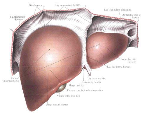
Human liver structure: distinguish convex upper diaphragmatic surface, facies diaphragmatica, inferior, locally concave, visceral surface, facies visceralis, acute lower edge, margo inferior separating front of upper and lower surfaces, and slightly convex posterior part, pars posterior. Diaphragm surface.
On the lower edge of the liver there is a notch of the round ligament, incisura ligaments teretis: to the right there is a small notch corresponding to the adjacent bottom of the gallbladder.
The diaphragmatic surface, facies diaphragmatica, is convex and corresponds to the shape of the dome of the diaphragm. From the highest point is a gentle slope to the lower sharp edge and to the left, to the left edge of the liver; A steep slope follows the back and right parts of the diaphragm surface. Above, to the diaphragm, there is a sagittally located peritoneal crescent ligament of the liver, lig. Falciforme hepatis, which follows from the lower edge of the liver back over approximately 2/3 of the width of the liver: behind the ligament leaves diverge to the right and left, passing into the coronary ligament of the liver, lig. Coronarium hepatis. The crescent ligament divides the liver, respectively, its upper surface into two parts: the right lobe of the liver, the lobus hepatis dexter, the largest and the largest, and the left lobe of the liver, the lobus hepatis sinister, the smaller. The upper part of the liver shows a slight cardiac impression, impressio cardiaca, formed as a result of the pressure of the heart and corresponding to the tendon center of the diaphragm.
On the diaphragmatic surface of the liver distinguish the upper part, pars superior, facing the tendon center of the diaphragm; Anterior part, pars anterior, facing anterior to the rib side of the diaphragm, and to the anterior wall of the abdomen in the epigastric region (left lobe); The right part, the pars dextra, directed to the right, to the lateral abdominal wall (respectively the middle axillary line), and the posterior part, pars posterior, facing the back.
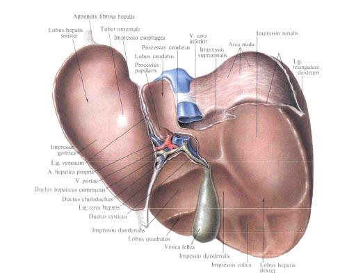
Visceral surface, facies visceralis, flat, slightly concave, corresponds to the configuration of the underlying organs. There are three furrows on it, dividing this surface into four parts. Two furrows are sagittal and extend almost parallel to each other from the anterior to posterior margin of the liver; Approximately at the middle of this distance, they are joined, as if in a crossbeam, by a third, transverse, furrow.
The left furrow consists of two sections: the anterior one, extending to the level of the transverse furrow, and the posterior one, located posteriorly from the transverse furrow. The deeper anterior section is the ligament of the circular ligament, fissura lig. Teretis (in the embryonic period - the furrow of the umbilical vein), begins at the lower edge of the liver from the incision of the circular ligament, incisura lig. Teretis. In it lies a round ligament of the liver, lig. Teres hepatis, going in front and below the navel and enclosing the obliterated umbilical vein. The posterior part of the left sulcus is the ligament of the venous ligament, fissura lig. Venosi (in the embryonic period - the fovea of the venous duct, fossa ductus venosi), contains a venous ligament, lig. Venosum (obliterated venous duct), and extends from the transverse furrow back to the left hepatic vein. The left furrow, according to its position on the visceral surface, corresponds to the attachment line of the crescent ligament on the diaphragmatic surface of the liver and thus serves as a boundary between the left and right lobes of the liver. At the same time, a round ligament of the liver is embedded in the lower edge of the crescent ligament, on its free anterior part.
The right sulcus is a longitudinally located fossa and is called the fossa of the gallbladder, fossa vesicae felleae, which corresponds to the cut on the lower edge of the liver. It is less deep than the groove of the circular ligament, but wider and represents the imprint of the gall bladder located in it, vesica fellea. The fossa extends posteriorly to the transverse furrow; The furrow of the inferior vena cava, sulcus venae cavae inferioris, extends posteriorly from the transverse furrow.
The transverse furrow is the portal of the liver, porta hepatis. It has its own hepatic artery, a. Hepatis propria, common hepatic duct, ductus hepaticus communis, and portal vein, v. Portae.
Both the artery and vein are divided into the main branches, right and left, already in the gates of the liver .

Three of these furrows divide the visceral surface of the liver into four lobes of the liver, lobi hepatis. The left furrow delimits the lower surface of the left lobe of the liver from the right; The right furrow delimits the lower surface of the right lobe of the liver from the left.
The middle section between the right and left furrows on the visceral surface of the liver is divided by a transverse furrow into the anterior and posterior grooves. The front portion is the square fraction, lobus quadratus, the posterior part is the caudate lobe, lobus caudatus.
On the visceral surface of the right lobe of the liver, closer to the anterior margin, there is a colon-intestinal impression, impressio colica; Behind, to the very posterior margin, are: to the right - a vast depression from the adjacent right kidney, a renal impression, impressio renalis, to the left - a duodenal-duodenal (duodenal) adjoining to the right furrow, impressio duodenalis; Even more posteriorly, to the left of the renal depression, - the right adrenal depression, adrenal depression, impressio suprarenalis.
The square fraction of the liver, lobus quadratus hepatis, is bounded to the right by the pit of the gallbladder, to the left - by the slit of the circular ligament, from the front by the lower edge, from the rear by the portal of the liver. In the middle of the width of the square share there is a depression in the form of a wide transverse groove - an imprint of the upper part of the duodenum , a duodenal intestinal depression, continuing here with the right lobe of the liver.
Hvostataya share of the liver, lobus caudatus hepatis, is located posteriorly from the gates of the liver, is confined to the front by a transverse groove of the gates of the liver, to the right by the furrow of the hollow vein, sulcus venae cavae, to the left by the slit of the venous ligament, fissura lig. Venosi, and behind - the back of the diaphragmatic surface of the liver. In the anterior part of the caudal lobe there is a small protrusion on the left - the papillary processus, processus papillaris, adjoining the posterior to the left side of the liver gates; On the right the caudate part forms the caudate processus, processus caudatus, which is directed to the right, forms a bridge between the posterior end of the pit of the gallbladder and the anterior end of the furrow of the inferior vena cava and passes to the right lobe of the liver.
The left lobe of the liver, lobus hepatis sinister, on the visceral surface, closer to the anterior margin, has a convexity - glandular tuber omentale, which faces the small omentum, omentum minus. On the posterior edge of the left lobe, directly next to the venous ligament slit, there is an impression from the adjacent abdominal part of the esophagus - the esophageal impression, impressio esophageale.
To the left of these formations, closer to the back, on the lower surface of the left Lobe there is a gastric impression, impressio gastrica.
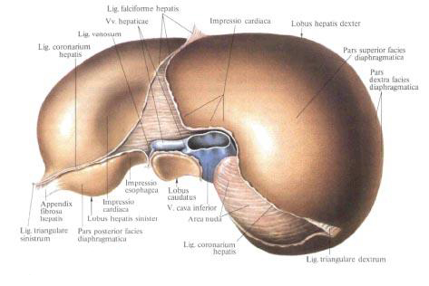
The posterior part of the diaphragmatic surface, pars posterior faciei diaphragmaticae, is a fairly wide, slightly rounded section of the surface of the liver. It forms a concavity corresponding to the place of attachment to the spine. Its central part is wide, and narrows to the right and to the left. Correspondingly, there is a groove in the right lobe, in which the lower hollow vein, the hollow vein groove, sulcus venae cavae is laid. Near the upper end of this furrow in the liver substance are visible three hepatic veins, venae hepaticae, flowing into the lower vena cava. The edges of the groove of the hollow vein are interconnected by a connective tissue bundle of the inferior vena cava.
The liver is almost completely surrounded by the peritoneum. The serosa, tunica serosa, covers its diaphragmatic, visceral surfaces and the lower edge. However, in areas where ligaments are suitable for the ligament and the gallbladder is adjacent, there remain areas of different widths that are not covered by the peritoneum. The largest non-covered abdominal area is on the back of the diaphragm surface, where the liver is directly attached to the posterior wall of the abdomen; It has the form of a rhombus-extraperitoneal field, area nuda. Accordingly, its greatest width is the lower hollow vein. The second such site is located in the location of the gallbladder. From the diaphragmatic and visceral surfaces of the liver, the peritoneal ligaments depart.
The structure of the liver.
The serous membrane, tunica serosa, covering the liver, is underlain by the subserous base, tela subserosa, and then by the fibrous membrane, tunica fibrosa. Through the gates of the liver and the posterior end of the ligament of the circular ligament, the connective tissue penetrates into the parenchyma in the form of a so-called perivascular fibrous capsule, capsula fibrosa perivascularis, in the processes of which there are bile ducts , branches of the portal vein and its own hepatic artery; In the course of the vessels it reaches from inside the fibrous membrane. Thus, a connective tissue framework is formed, in the cells of which there are hepatic lobules.
Lobule of the liver.
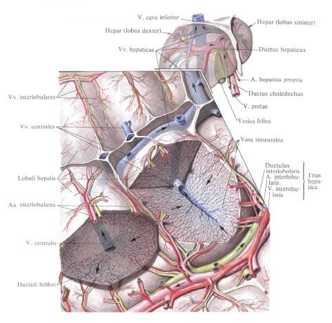
Lobule of the liver , lobulus hepaticus, 1-2 mm in size. Consists of hepatic cells - hepatocytes, hepatocyti, forming hepatic plates, laminae hepaticae. In the center of the lobule is the central vein, v. Centralis, and around the lobule are interlobular arteries and veins, aa. Interlobular et vv, interlobulares, from which originate interlobular capillaries, vasa capillaria interlobularia. Interlobular capillaries enter the lobule and pass into the sinusoid vessels, vasa sinusoidea, located between the hepatic plates. In these vessels the arterial and venous (from v, portae) blood is mixed. Sinusoid vessels flow into the central vein. Each central vein is poured into podolkovye, or collective, veins, vv. Sublobulares, and the latter - in the right, middle and left hepatic veins. Vv. Hepaticae dextrae, mediae et sinistrae.
Between the hepatocytes bile ducts, canaliculi biliferi, which enter the bile ducts , ductuli biliferi, and the latter outside the lobules connect to the interlobular bile ducts , ductus interlobulares biliferi. Segmental ducts are formed from the interlobular bile ducts.
Based on the study of intrahepatic vessels and bile ducts There was a modern understanding of the shares, sectors and segments of the liver. Branches of a portal vein of the first order bring blood to the right and left lobes of the liver, the boundary between which does not correspond to the external border, but passes through the pit of the gallbladder and the furrow of the inferior vena cava.
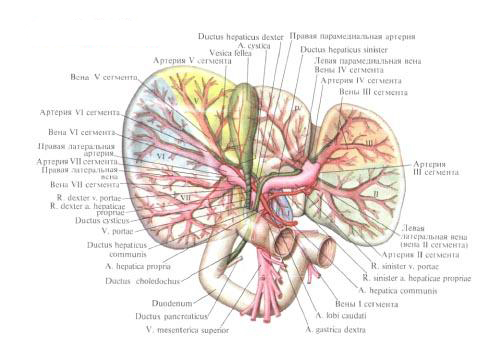
Branches of the second order provide the inflow of blood to the sectors: in the right lobe - in the right pyramedian sector, sector paramedianum dexter, and right lateral sector, sector lateralis dexter; In the left lobe - in the left paramedian sector, sector paramedianum sinister, left lateral sector, sector lateralis sinister, and left dorsal sector, sector dorsalis sinister. The last two sectors correspond to the I and II segments of the liver. Other sectors are divided into two segments, so that in the right and left segments of 4 segments.
Lobes and segments of the liver have their bile ducts, branches of the portal vein and their own hepatic artery. The right lobe of the liver is drained by the right hepatic duct, the ductus hepaticus dexter, which has the anterior and posterior branches, r. Anterior et r. Posterior, left part of liver - left hepatic duct, ductus hepaticus sinister, consisting of medial and lateral branches, r. Medialis et lateralis, and the caudate part - by the right and left ducts of the caudate lobe, ductus lobi caudati dexter and ductus lobi caudati sinister.
The anterior branch of the right hepatic duct is formed from the ducts of segments V and VIII; The posterior branch of the right hepatic duct - from the ducts of segments VI and VII; The lateral branch of the left hepatic duct - from the ducts of segments II and III. Protocols of the square lobe of the liver flow into the medial branch of the left hepatic duct - the duct of segment IV, and the right and left ducts of the caudate lobe, the ducts of the I segment, can flow together Or separately into the right, left and common hepatic ducts, as well as into the posterior branch of the right and lateral branches of the left hepatic ducts. There may be other variants of the connection of I-VIII segmental ducts. The channels of the III and IV segments are often connected together.
The right and left hepatic ducts at the anterior edge of the liver gate or already in the hepatic-duodenal ligament form a common hepatic duct, ductus hepaticus communis.
The right and left hepatic ducts and their segmental branches are not permanent formations; If they are absent, the ducts that form them flow into the common hepatic duct. The length of the common hepatic duct is 4-5 cm, its diameter is 4-5 cm. The mucous membrane is smooth and does not form folds.
Topography of the liver.
Topography of the liver. The liver is located in the right subcostal area, in the epigastric region and partly in the left hypochondrium region. Skeleototypically, the liver is defined by projection onto the breast walls. On the right and in front along the middle clavicular line the highest point of the liver position (right lobe) is determined at the level of the fourth intercostal space; To the left of the sternum the highest point (left lobe) is at the level of the fifth intercostal space. The lower edge of the liver on the right along the middle axillary line is determined at the level of the tenth intercostal space; Further forward the lower border of the liver follows along the right half of the costal arch. At the level of the right sredneklyuchichnoy line, it comes out from under the arc, goes from right to left and up, crossing the epigastric region. The white line of the abdomen, the lower edge of the liver crosses in the middle of the distance between the xiphoid process and the umbilical ring. Further at level VIII of the left costal cartilage, the lower border of the left lobe crosses the costal arch to meet the upper border to the left of the sternum.
Behind the right, along the scapular line, the border of the liver is defined between the seventh intercostal (or VIII rib) at the top and the top edge of the XI rib at the bottom.
Synopia of the liver. At the top, the upper part of the diaphragmatic surface of the liver adjoins the right and partially to the left dome of the diaphragm , in front of it the anterior part adjoins in sequence to the rib side of the diaphragm and to the anterior abdominal wall: behind the liver adjoins the X and XI thoracic vertebrae and diaphragm legs, abdominal esophagus , And to the right adrenal. The visceral surface of the liver adjoins the cardiac part, the body and the pylorus, to the upper part of the duodenum , the right kidney, the right bend of the colon and to the right end of the transverse colon . The gallbladder is also adjacent to the inner surface of the right lobe of the liver.
You will be interested to read this:

Comments
When commenting on, remember that the content and tone of your message can hurt the feelings of real people, show respect and tolerance to your interlocutors even if you do not share their opinion, your behavior in the conditions of freedom of expression and anonymity provided by the Internet, changes Not only virtual, but also the real world. All comments are hidden from the index, spam is controlled.