


The gall bladder. Bile ducts
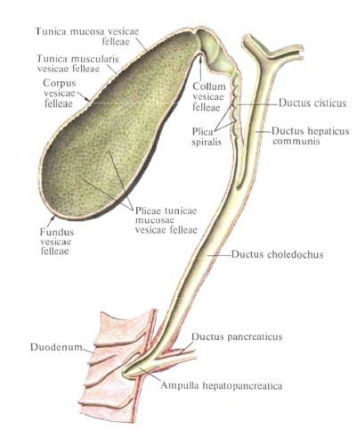
The gall bladder , vesica fellea (biliaris), is a sacciform reservoir for the bile produced in the liver; It has an elongated shape with a wide and narrow ends, and the width of the bladder from the bottom to the neck decreases gradually. The length of the gallbladder varies from 8 to 14 cm, the width is 3-5 cm, the capacity reaches 40-70 cm 3 . It has a dark green color and a relatively thin wall.
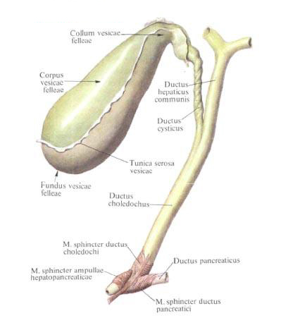
In the gallbladder, the bottom of the gallbladder is distinguished, the fundus vesicae felleae, the most distal and wide part of it, the body of the gallbladder, corpus vesicae felleae, the middle part and the cervix of the gall bladder, the collum vesicae felleae, the proximal narrow part from which the vesicle , Ductus cysticus. The latter, having united with the common hepatic duct, forms the common bile duct, ductus choledochus.
The gallbladder lies on the visceral surface of the liver in the fossa of the gallbladder, fossa vesicae felleae, separating the anterior section of the right lobe from the square lobe of the liver. The bottom of it is directed forward to the lower edge of the liver in the place where a small notch is located, and protrudes from under it; The cervix is turned towards the gates of the liver and lies together with the vesicular duct in the duplication of the hepatic duodenal ligament. At the junction of the body of the gallbladder, a bend usually forms in the neck, so the neck turns out to be at an angle to the body.
The gallbladder, located in the pit of the gallbladder, adjoins it with its upper, devoid of peritoneal surface and connects with the fibrous membrane of the liver. Its free surface, turned downwards, into the abdominal cavity, is covered with a serous leaf of the visceral peritoneum, passing to the bladder from the adjacent areas of the liver. The gallbladder can be located intraperitoneally and even have a mesentery. Usually, the bottom of the bladder protruding from the liver scissor is covered with the peritoneum on all sides.
The structure of the gallbladder.
The structure of the gallbladder. The wall of the gallbladder consists of three layers (with the exception of the upper extraperitoneal wall): the serosa, tunica serosa vesicae felleae, the muscular membrane, tunica muscularis vesicae felleae, and the mucosa, tunica mucosa vesicae felleae. Under the peritoneal wall of the bladder covers a thin loose layer of connective tissue - the subserous base of the gallbladder, tela subserosa vesicae felleae; On the extraperitoneal surface it is more developed.
The muscular membrane of the gallbladder, tunica muscularis vesicae felleae, is formed by a single circular layer of smooth muscles, among which there are also bundles of longitudinally and obliquely arranged fibers. The muscular layer is less pronounced in the region of the bottom and stronger - in the neck region, where it directly passes into the muscular layer of the cystic duct.
The mucous membrane of the gallbladder, tunica mucosa vesicae felleae, is thin and forms numerous folds, plicae tunicae mucosae vesicae felleae, giving it the appearance of a network. In the neck area, the mucosa forms several oblique spiral folds one after the other, plicae spirales. The mucous membrane of the gallbladder is lined with a single-row epithelium; In the region of the neck in the submucosa base there are glands.
Topography of the gallbladder.
Topography of the gallbladder. The bottom of the gallbladder is projected on the anterior abdominal wall in the corner formed by the lateral margin of the right rectus abdominis and the edge of the right costal arch, which corresponds to the end of the IX rib cartilage. Synotypically, the lower surface of the gallbladder is adjacent to the anterior wall of the upper part of the duodenum; To the right of it adjoins the right bend of the colon.
Often, the gallbladder is connected to the duodenum or to the colon with the peritoneal fold.
Blood supply: from the gall-bladder artery, a. Cystica, branches of the hepatic artery.
Bile ducts.
Extrahepatic bile ducts are three: a common hepatic duct, ductus hepaticus communis, a ductus ductus, ductus cysticus, and a common bile duct, ductus choledochus (biliaris).
The common hepatic duct, ductus hepaticus communis, is formed in the gates of the liver as a result of the fusion of the right and left hepatic ducts, the ductus hepaticus dexter and sinister, the latter are formed from the intrahepatic ducts described above. Having descended into the hepatic-duodenal ligament, the common hepatic duct connects with the vesicular duct Duct flowing from the gallbladder; Thus there is a common bile duct, ductus choledochus.
The bladder duct, ductus cysticus, has a length of about 3 cm, its diameter is 3-4 mm; The neck of the bladder forms with the body of the bladder and with the bladder duct two bends. Then, in the hepatic-duodenal ligament, the duct is directed from the top to the right downwards and slightly to the left and usually merges at an acute angle with the common hepatic duct. The muscular membrane of the vesicular duct is poorly developed, although it contains two layers: longitudinal and circular. Over the course of the cystic duct, its mucous membrane forms a spiral fold a few turns, plica spiralis.
Common bile duct, ductus choledochus. Is incorporated in the hepatic-duodenal ligament. It is a direct continuation of the common hepatic duct. Its length is on the average 7-8 cm, sometimes reaches 12 cm. There are four sections of the common bile duct:
- Located above the duodenum;
- The back of the upper part of the duodenum;
- It is located between the head of the pancreas and the wall of the descending part of the intestine;
- Adjacent to the head of the pancreas and passing obliquely through it to the wall of the duodenum.
The wall of the common bile duct, unlike the wall of the common hepatic and cystic ducts, has a more pronounced muscular membrane that forms two layers: longitudinal and circular. At a distance of 8-10 mm from the end of the duct, the circular muscular layer is thickened, forming a common bile duct sphincter, m. Sphincter ductus choledochi. The mucous membrane of the common bile duct of the folds does not form, with the exception of the distal portion, where there are several folds. In the submucosa-based walls in non-hepatic bile ducts there are mucous glands of the bile ducts, glandulae mucosae biliosae.
The common bile duct connects to the pancreatic duct and flows into the common cavity - the hepatic-pancreatic ampulla, ampulla hepatopancreatica, which opens into the lumen of the descending part of the duodenum at the apex of its large papilla, papilla duodeni major, 15 cm from the pylorus. The size of the ampoule can reach 5-12 mm.
The type of confluence of the ducts can vary: they can open into the intestine by separate mouths or one of them may flow into another.
In the area of the large papilla of the duodenum, the mouth of the ducts are surrounded by a muscle - this is the sphincter of the hepatic pancreas ampulla (sphincter of the ampulla), m. Sphincter ampullae hepatopancreaticae (m. Sphincter ampulae). In addition to the circular and longitudinal layers, there are separate muscle bundles forming an oblique layer that unites the ampulla sphincter with the sphincter of the common bile duct and with the sphincter of the pancreatic duct.
Topography of bile ducts. Extrahepatic ducts are laid in the hepatic-duodenal ligament together with the common hepatic artery, its branches and portal vein. The common bile duct is located at the right edge of the ligament, to the left of it is the common hepatic artery, and deeper than these formations and between them - the portal vein; In addition, lymphatic vessels, nodes and nerves lie between the ligament sheets.
The division of the hepatic artery into the right and left hepatic branches occurs in the middle of the ligament length, the right hepatic branch going upward passes under the common hepatic duct; In the place of their intersection from the right hepatic branch, the gall-bladder artery, a. Cystica, which is directed to the right and upward into the region of the angle (gap) formed by the fusion of the vesicular duct with the common hepatic duct. Further, the gallbladder artery runs along the wall of the gallbladder.
Innervation: liver, gallbladder and bile ducts - plexus hepaticus (truncus sympathicus, nn. Vagi).
Blood supply: liver - a. Hepatica propria, and its branch a. The cystica approaches the gallbladder and its ducts. In addition to the artery, v. Enters the gates of the liver. Portae, which collects blood from unpaired organs in the abdominal cavity; After passing through the system of intraorganic veins, leaves the liver through vv. Hepaticae. Flowing into v. Cava inferior. From the gallbladder and its ducts venous blood flows into the portal vein. Lymph is removed from the liver and gall bladder in nodi lymphatici hepatici, phrenici superior and inferior, lumbales dextra, celiaci, gastrici, pylorici, pancreatoduodenales, anulus lymphaticus cardiae, parasternales.
You will be interested to read this:

Comments
When commenting on, remember that the content and tone of your message can hurt the feelings of real people, show respect and tolerance to your interlocutors even if you do not share their opinion, your behavior in the conditions of freedom of expression and anonymity provided by the Internet, changes Not only virtual, but also the real world. All comments are hidden from the index, spam is controlled.