


Osteology - the doctrine of bones
Torso bones
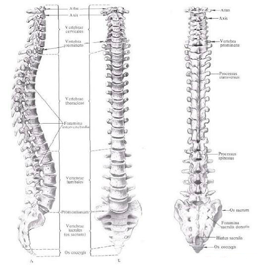
Vertebral column
Chest bones
Bones of the head
Skull bones
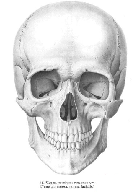
Face Bones
- Lower nasal concha
- Nasal bone
- Lacrimal bone
- Vomer
- Upper jaw
- The palatine bone
- Cheekbone
- Lower jaw
- The hyoid bone
Skull as a whole
- Skull Skull
- Spring
- Base of skull
- The nasal cavity
- Bony sky
- Glaznitsa
- Temporal, transverse and pterygopalatine fossa
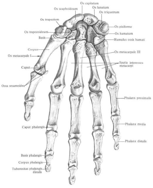
Bones of upper limb
Belt of the upper limb
Free part of upper limb
Bones of lower limb
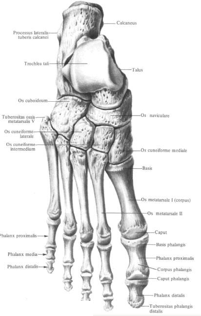
Belt of lower limb (pelvic girdle)
Free part of the lower limb
Bones, ossa , are a solid support of the soft tissues of the body and form levers that move by force of muscle contraction. In the body of the bone form a system of the skeleton, the systema skeletale , which includes the axial skeleton, skeleton axiale , and an additional skeleton, skeleton appendiculare . The axial skeleton includes the skull, cranium , vertebral column, columna vertebralis , and the bones of the chest, ossa thoracis . The additional skeleton combines the bones of the upper limb, ossa membri superioris , and the bones of the lower limb ossa membri inferioris .
The skeleton system includes more than 200 bones, 85 of them are paired.

Each bone is an organ constructed from various types of connective tissue, containing a bone marrow, equipped with vessels and nerves,
In the skeleton system, the osseous part, pars ossea , and the cartilaginous part, pars cartilaginosa , are distinguished . The main part in the system of the skeleton is the bone part. The cartilaginous part of the skeleton system includes articular cartilages, cartilagines articulares , epiphyseal cartilages, cartilagines epiphysiales , and costal cartilages , cartilagines costales .
Outside, the bone is covered by a thin connective tissue membrane - the periosteum, periosteum , in which the fibrous and osteogenic layers are distinguished. The superficially located fibrous layer connects to the bone penetrating into it fibers (perforating fibers), contains blood and lymph vessels, nerves. Hence the vessels and nerves pass into the bone through the nutrient holes, foramina nutricia , and further through the nutritional canalis canalis nutricius, into the bone marrow. The inner, osteogenic, layer includes the forming cells (osteoblasts) involved in the processes of development and reconstruction of bone tissue, including after traumas and fractures. On the border with the articular cartilage covering the ends of the bone, the periosteum passes into the perichondrium, perichondrium, As a result, the bone turns out to be enveloped by a continuous connective tissue membrane. This shell covers the surface of the bone and all the formations on it: processes , processus ; Awn , spinae ; Cristae , cristae ; Hillocks , tuberi ; Tuberculi , tuberculi ; Rough lines, liniae asperae ; Deepening, foveae ; Fossae , fossae , etc.
Thinner shell - endost, endosteum , - lining the bone from the inside.
In shape, the bones are long, ossa longi , short, ossa brevia , and flat, ossa plana . Some bones inside have air-filled cavities; Such bones are called airborne, ossa pneumatica . In addition, some bones are referred to abnormal (mixed) bones, ossa irregularea , which consist of suits having different shapes and structures .
In the long bones (humerus, collarbone , metacarpal , phalanges , etc.), the middle part is distinguished - diaphysis , diaphysis , and the two terminal sections - epiphyses , epiphyses . Epiphysis, located closer to the axial skeleton. Called proximal, epiphysis proximalis , and the epiphysis of the same bone, occupying a position farther from the axial skeleton, is distal, epiphysis distalis .

Sites of long bones located on the border of the diaphysis and epiphyses are called metaphyses, the border itself is visible only in the bones of children and adolescents, while the interlayer of cartilage-epiphyseal cartilage, cartilago epiphysialis- remains between the diaphysis and the epiphyses . Due to this cartilage, the bone grows intensively in length. Then the epiphyseal cartilage is replaced by a bone tissue forming the epiphyseal line, linea epiphysialis , which with age becomes almost indistinguishable.
On the long bone cut can be distinguished a compact substance, the substantia compacta , which forms the outer layers of the bone, and the spongy (trabecular) substance , substantia spongiosa , located inside of the compact substance. Mainly in the epiphyses and metaphyses. In the diaphysis of long bones, a compact substance surrounds the mediastinal cavitas cavitas medullaris , which has the shape of a tube.
In short bones, a thin layer of a compact substance is defined on the cut at the surface, surrounding the sponges of the spongy substance, which forms the bulk of the bone. Trabeculae spongy matter form a complex mesh network. They are located in each bone in strict accordance with its functional loads.
In flat bones, on the contrary, the spongy substance usually forms a thin layer of bone and is surrounded on both sides by plates of a compact substance. In the bones of the cranial vault, the spongy substance is called diploe, diploe (double) ; It lies between the outer and inner plates of a compact substance, laminae externa et interna . In the thickness of the spongy substance of the bones of the cranial vault, diplic canals pass, canales diploici, conducting venous vessels.
Some bones of the skull (frontal, latticed, wedge-shaped, upper jaw) contain airborne sinuses communicating with the nasal cavity. At the same time, a number of sections of the bones of the skull form thickenings - buttresses (frontal-nasal, alveolar-malar, pterygoid, mandibular) Which are the base places of these bones. Thanks to the buttresses, the mechanical tremors experienced by the skull are relaxed.
In the cells of the spongy substance of the bone and in the medullary cavity the bone marrow, medulla ossium , is contained . There are red bone marrow, medulla ossium rubra , and yellow bone marrow, medulla ossium flava .
Red bone marrow has high functional activity and is able to form blood elements. As the body develops and grows, the red bone marrow gradually becomes yellow. The yellow bone marrow is less active, performs a reserve role, but can be activated under certain conditions, partially turning into red.

Comments
When commenting on, remember that the content and tone of your message can hurt the feelings of real people, show respect and tolerance to your interlocutors even if you do not share their opinion, your behavior in the conditions of freedom of expression and anonymity provided by the Internet, changes Not only virtual, but also the real world. All comments are hidden from the index, spam is controlled.