


Bundles of the spine, functions, structure
Vertebral ligaments , ligg. Columnae vertebralis, can be divided into long and short.

The group of long ligaments of the vertebral column includes the following:
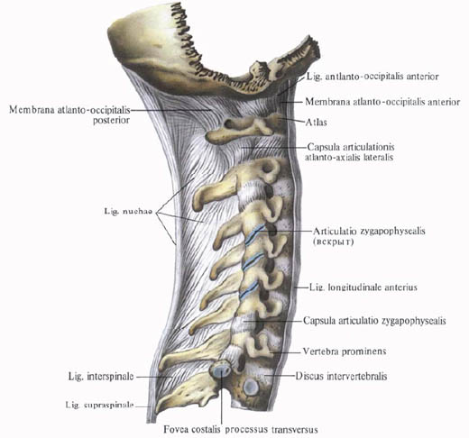
1. Anterior longitudinal ligament, lig. Longitudinale anterius, extends along the anterior surface and partly along the lateral surfaces of the vertebral bodies from the anterior tubercle of the atlant to the sacrum, where it is lost in the periosteum of the I and II sacral vertebrae.
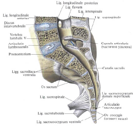
The anterior longitudinal ligament in the lower parts of the spine is much wider and stronger. It loosely combines with the bodies of the vertebrae and tightly - with the intervertebral discs, since it is woven into the perichondrium covering them, perichondrium; No lateral vertebrae it extends into their periosteum. The deep layers of the bundles of this bundle are somewhat shorter than the surface ones, so that they connect the adjacent vertebrae with each other, and the superficial, longer beams lie over 4 vertebrae. The anterior longitudinal ligament limits the excessive extension of the vertebral column.
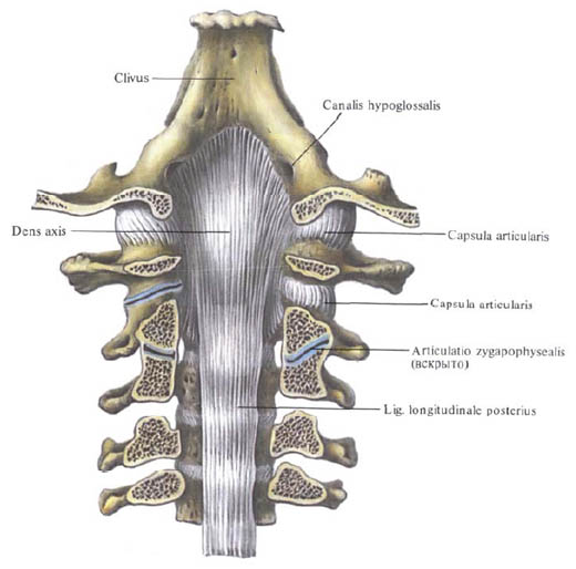
2. The posterior longitudinal ligament, lig, longitudinale posterius, is located on the posterior surface of vertebral bodies in the vertebral canal. It originates on the posterior surface of the axial vertebra, and at the level of the two upper cervical vertebrae it extends into the cover membrane, the tetraplea tectoria. The ligament reaches the initial section of the sacral canal. The posterior longitudinal ligament, in contrast to the anterior one in the upper part of the spinal column, is wider than in the lower one. It is firmly fused to the intervertebral discs, at the level of which it is somewhat wider than at the level of the vertebral bodies. With the bodies of the vertebrae, it connects loose, and in the layer of connective tissue between the ligament and the body of the vertebra lies the venous plexus. The superficial bundles of this ligament, as well as the anterior longitudinal ligament, are longer than the deep ones.
The group of short ligaments of the spine is syndesmosis.
These include the following links:

1. Yellow ligament, ligg. Flava, perform gaps between the arcs of the vertebrae from the axial vertebra to the sacrum. They are directed from the inner surface and the lower edge of the arch of the overlying vertebra to the outer surface and the upper edge of the arch of the underlying vertebra and their front edges border the intervertebral openings.
The yellow bundles consist of vertically extending elastic beams, giving them a yellow color. They reach the greatest development in the lumbar region. The yellow ligaments are very elastic and elastic, so when the trunk is unbent, they shorten and act like muscles, causing the trunk to keep in the state of extension and reducing the muscle tension. When bending, the ligaments are stretched and thus also reduce the tension of the trunk rectifier (see "Muscles of the back"). There are no yellow bands between the arcs of the atlas and the axial vertebra. Here, the cover membrane is stretched, which, by its front edge, delimits from behind the intervertebral opening, through which the second cervical nerve emerges.
2. Interstitial ligament, ligg. Interspinalia, - thin laminae, performing the intervals between the spinous processes of two adjacent vertebrae. They reach the highest power in the lumbar spine and are least developed between the cervical vertebrae. In front are connected with yellow ligaments, and behind, at the apex of the spinous process, merge with the bony bundle.
3. A ligamentous ligament, tig.supraspinale, is a continuous cord that runs along the apices of the spinous processes of the vertebrae in the lumbar and thoracic areas. At the bottom, it is lost on the spinous processes of the sacral vertebrae, at the top at the level of the protruding vertebra (CVII) passes into the rudimentary ligament ligament.
4. A ligamentous ligament. Lig. Nuchae, - a fossil plate consisting of elastic and connective tissue bundles. It is directed from the spinous process of the protruding vertebra (CVII) along the spinous processes of the cervical vertebrae upward and, somewhat widening, is attached to the external occipital crest and the external occipital protrusion; Has the form of a triangle.
5. Cross-connective ligaments, Iigg. Intertransversaria, are thin beams, slightly expressed in the cervical and partly thoracic areas and more developed in the lumbar region. This pair of ligaments, connecting the tips of the transverse processes of adjacent vertebrae, limit lateral movements of the spine in the opposite direction. In the cervical region, they can be bifurcated or absent.
You will be interested to read this:

Comments
When commenting on, remember that the content and tone of your message can hurt the feelings of real people, show respect and tolerance to your interlocutors even if you do not share their opinion, your behavior in the conditions of freedom of expression and anonymity provided by the Internet, changes Not only virtual, but also the real world. All comments are hidden from the index, spam is controlled.