
In the Netherlands, created a 3D atlas of embryonic development

An embryo (another Greek) or an embryo is an early stage in the development of an animal and a person, starting from a fertilized egg ( zygote ). The embryonic development, which usually occurs in the egg shells or special organs of the mother's body, is completed by the appearance of the ability to self-feed and actively move.
The Dutch scientists created a three-dimensional atlas of prenatal development of a man in the first two months after conception . This is reported by 3D Embryology .
Embryonic development
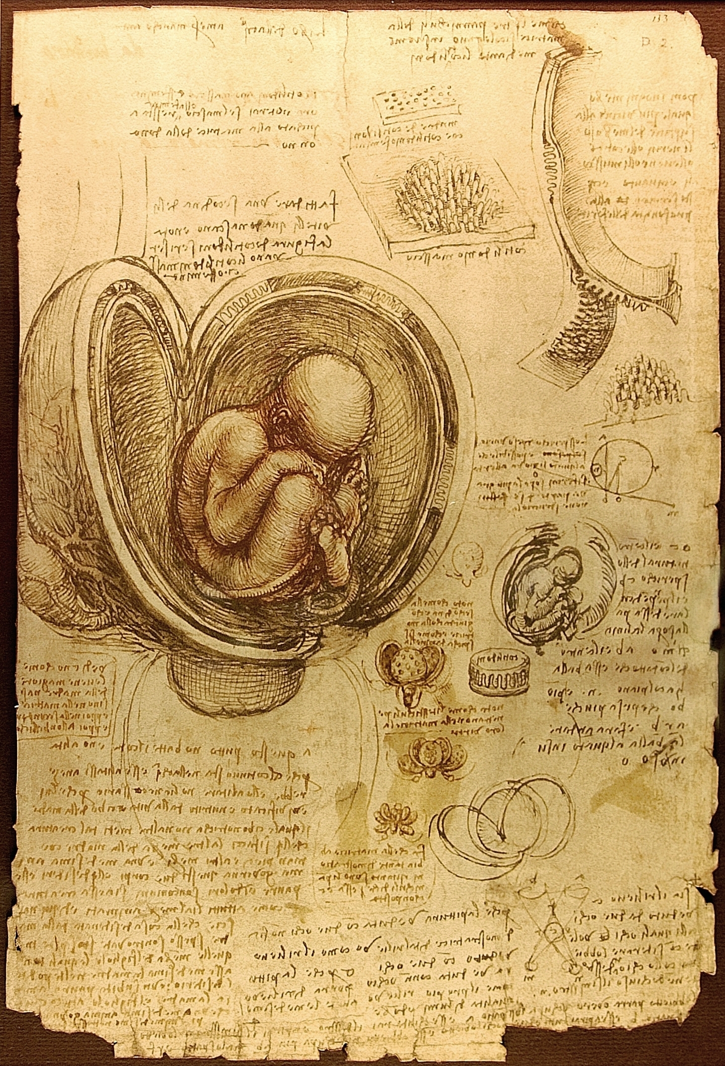
The human embryo. Picture of Leonardo da Vinci (about 1510-1513)
Embryonic, or embryonic, development of a living organism occurs either in the egg shells outside the mother's body, or inside it.
In the course of this development, a multicellular organism, consisting of various organs and tissues that is capable of independent existence, emerges from the ovule. In all animals, embryonic development involves the fertilization of the egg, or, in the case of parthenogenesis, its activation, followed by the stages of crushing, gastrulation, organogenesis, followed by emergence from the egg shells or birth.
Fertilization occurs either in the mother's body or in the aquatic environment. After fertilization, the crushing of the egg follows, during which it is consistently and repeatedly divided into blastomeres - first large ones, and then more and more small cells. As a result, there is a multicellular organism - a blastula. The chain of cleavage divisions creates the prerequisites for the emergence of differentiation, that is, the differences between parts of the embryo. The primary differentiation is due to the unequal composition of the cytoplasm of cells that originated from different parts of the egg. The ability of embryonic cells to move is also important for the formation of adult organs.
At the stage of gastrulation, the embryonic sheets are separated, and as a result, a three-layer structure emerges: the ectoderm (outer layer), the endoderm (inner layer), the mesoderm (intermediate layer).
Although in the early stages of development embryonic cells can develop in different directions, under the influence of a number of factors they gradually determine (acquire the ability to develop in only one specific direction).
At the stage of organogenesis, which is provided mainly by a variety of cellular movements and differentiation of the cells themselves, there is a division of the embryonic sheets into the rudiments of organs and systems, during which large germs differentiate into smaller ones, and as a result an increasingly complex structure of the whole organism . For example, from the part of the ectoderm that forms the rudiment of the nervous system, the brain is allocated. From the latter, the rudiments of the eyes are distinguished, in which the retina is allocated, and in it the specialized visual cells-rods and cones are differentiated. The embryonic development of various groups of animals is not the same: a large yolk sac develops in the embryos of fish, the yolk sac and the special organs - the allantois and amnion - are peculiar to birds, and the mammal also has a trophoblast and placenta.
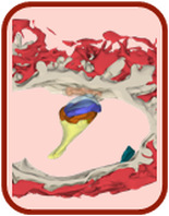
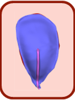
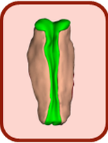
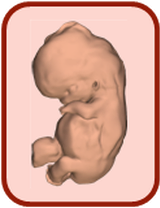
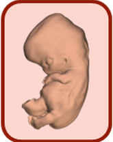
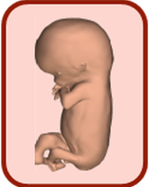
3D atlas of embryonic development
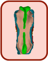
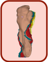
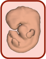
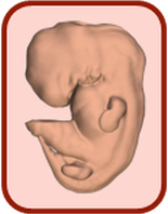
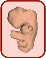
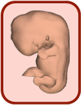
Scientists of the Academic Medical Center in Amsterdam identified and labeled 150 structures and organs of the body of embryos, after which their three-dimensional computer models were reconstructed.
The results of the studies were grouped into a single atlas of embryo development in the period from 15-17 to 56-60 days after conception.
All models were classified into development stages and converted into an interactive 3D-PDF format, which is freely available.

You can download the models from the project website. The level of development of the embryo is divided into stages, switching to which you can see the model and its individual parts.

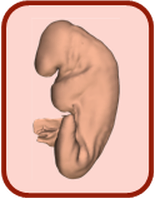
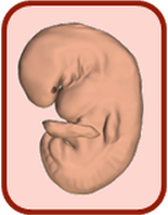
The Atlas allows us to consider both the embryo as a whole, and its individual structures, organs and systems.

For a more complete understanding, scientists indicated the size of the embryo in millimeters and compared with the palm of an adult.
As noted, work on the creation of the atlas was conducted since 2009. Participation in it was accepted by a group of embryologists with the assistance of 75 students.
3D-PDF's
Stage 7 (15-17 days) Specimen 8752
Stage 8 (17-19 days) Specimen 8671
Stage 9 (19-21 days) Specimen H712
Stage 10 (21-23 days) Specimen 6330
Stage 11 (23-26 days) Specimen 6784
Stage 12 (26-30 days) Specimen 8505A
Stage 13 (28-32 days) Specimen 836
Stage 15 (35-38 days) Specimen 3512
Stage 16 (37-42 days) Specimen 6517
Stage 17 (42-44 days) Specimen 6521
Stage 18 (44-48 days) Specimen 6524
Stage 20 (51-53 days) Specimen 462
Stage 21 (53-54 days) Specimen 7254
Stage 23 (56-60 days) Specimen 9226

Download All: http://3datlas.3dembryo.nl/3Datlas_pdf_all_v2016-03.zip (~ 84 MB)
3D Atlas of Human Embryology - data
- Date: 24 November 2016
- Contact: [email protected]
- Site: http://3datlas.3dembryo.nl/
This website is for downloading data of the 3D Atlas.
Microphotographs of specimens of the Carnegie Collection are available with permission of the caretakers of this collection.
3D-PDF files: Download the 3D-PDFs and use Adobe Reader X (or newer) on MS Windows or Mac OS for 3D interaction.
3D-pdf files: https://get.adobe.com/reader Download the 3D-pdf's and use Adobe Reader X (or newer) on MS Windows or Mac OS for 3D interaction.
3D-tiff files: http://www.irfanview.com IrfanView can be used for easy viewing of multipage tiff files. Use CTRL + PageDown / PageUp for scrolling trough the gray and label stack.
Amira files: http://www.amira.com/ Use Amira for combined use of the gray and label files. The label files also contain the labelnames.
Via 3dembryoatlas.com & hromadske.ua & wiki

Comments
When commenting on, remember that the content and tone of your message can hurt the feelings of real people, show respect and tolerance to your interlocutors even if you do not share their opinion, your behavior in the conditions of freedom of expression and anonymity provided by the Internet, changes Not only virtual, but also the real world. All comments are hidden from the index, spam is controlled.