Occipital bone

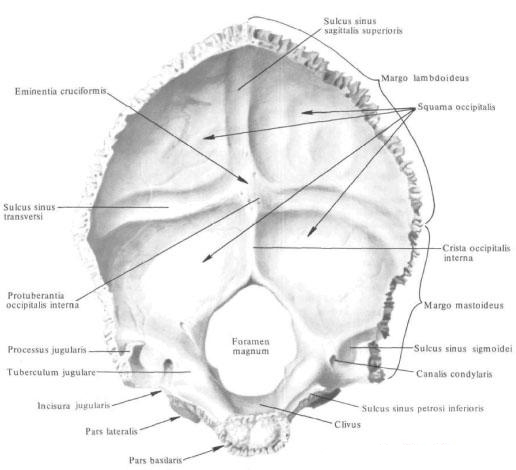
The occipital bone , os occipitale, is unpaired, forms the posterior part of the skull. Its outer surface is convex, but its inner, cerebral, concave. In the anterior section of the department there is a large (occipital) opening, foramen magnum, connecting the cranial cavity with the vertebral canal. This hole is surrounded by a shallow furrow of the occipital sinus, sulcus sinus occipitalis. Based on the data on the process of development of the occipital bone, it distinguishes four parts surrounding the large (occipital) opening: the basilar part - in front of the large (occipital) opening, the paired lateral parts - on each side of it and the occipital scales located behind.
Basilar part , pars basilaris. Short, thick, quadrangular; Its posterior margin free, smooth and slightly pointed, bordering the front with a large (occipital) opening; Anterior edge thickened and rough, connected to the body of the sphenoid bone by means of cartilage, forming a wedge-occipital synchondrosis, synchondrosis sphenooccipitalis.
In adolescence, cartilage is replaced with bone tissue and both bones merge into one. The upper surface of the basilar part, facing the cavity of the skull , is smooth and slightly concave. It consists of a stingray located in front of it, a clivus directed towards the large (occipital) opening (on it lie the medulla oblongata, the bridge and the basilar artery of the brain with branches). In the middle of the lower, outer, slightly convex surface of the basilar part there is a small pharyngeal tubercle, tuberculum pharyngeum (place of attachment of the anterior longitudinal ligament and pharyngeal membrane of the pharynx), and rough lines (traces of attachment of the straight anterior and long muscles of the head).
The outer, slightly uneven edge of the basilar part and lateral parts of the occipital bone adjoins the posterior edge of the stony part of the temporal bone . Between them a stony-occipital fissure is formed, fissura petrooccipitalis; On an uncertified skull it is made of cartilage, forming a stony-occipital synchondrosis, synchondrosis petrooccipitalis, which as a rest of the cartilaginous skull ossifies with age.
Lateral parts, paries laterales, somewhat elongated, in the posterior sections thickened, and in the anterior ones somewhat narrowed; They form the lateral sides of the large (occipital) aperture, fused in front with the basilar part, and behind - with the occipital scales.

On the cerebral surface of the lateral part, at its outer edge, there is a narrow groove of the lower stony sinus, sulcus sinus petrosi inferioris, which adjoins the posterior edge of the stony part of the temporal bone , forming a channel with the epithelial furrow of the temporal bone, where the venous lower stony sinus, sinus petrosus Inferior.
On the lower, outer surface of each lateral part is an oblong-oval shaped convex articular process - occipital condyles, condylus occipitalis. Their articular surfaces come closer in front, they diverge from behind; They articulate with the upper articular pits of the atlas. Behind the occipital condyle is the condylar fossa, fossa condylaris, and at the bottom - an opening leading to the unstable condyle canal, canalis condylaris, which is the site of the condyle of the condyle emissary vein, v. Emissaria condylaris.
On the outer edge of the lateral part there is a large, with smooth edges, the jugular notch, incisura jugularis, on which protrudes a small intragenic process, processus intrajugularis. Jugular tenderloin with the same name of the pit of the stony part of the temporal bone forms a jugular hole, foramen jugulare.
The internuclear processes of both bones divide this opening into two parts: the large posterior one, in which the upper bulb of the internal jugular vein lies, bulbus v. Jugularis superior, and a smaller anterior, through which the cranial nerves pass: glossopharyngeal, n. Glossopharyngeus, wandering, n. Vagus, and additional, n. Accessorius.
The jugular process, the processus jugularis, restrains the jugular cut from behind and outside. On the outer surface of its base there is a small parotid process, processus paramastoideus (the attachment site of the rectus lateral muscle of the head , M. rectus capitis lateralis).
Behind the jugular process, from the inner surface of the skull, is a wide sulcus sigmoid sulcus, sulcus sinus sigmoidei, which is the continuation of the epithelium of the same name with the temporal bone. A smooth jugular tubercle, tuberculum jugulare, lies anterior and medial. Behind and down from the jugular tubercle, between the jugular process and the occipital condyle, in the thickness of the bone passes the sublingual canal, canalis hypoglossalis (in it lies the sublingual nerve, n. Hypoglossus).
The occipital scales, squama occipitalis, confine a large (occipital) opening behind and make up the greater part of the occipital bone. It is a wide curved plate of triangular shape with a concave inner (cerebral) surface and a convex outer surface.
The lateral margin of the scales is divided into two sections: a larger upper, heavily serrated lambdoid margin, margo lambdoideus, which, joining the occipital margin of the parietal bones, forms a lambdoid suture, sutura lambdoidea, and a smaller lower, slightly serrate mastoid margin, margo mastoideus , Adjacent to the edge of the mastoid process of the temporal bone, forms the occipital mastoid suture, sutura occipitomastoidea.
In the middle of the outer surface of the scales, in the region of its greatest prominence, there is an external occipital protuberance, protuberantia occipitalis externa, easily palpable through the skin. From it diverge in pairs paired convex upper fringes, lineae nuchae superiores, above which and parallel to them there are additional highest fetal lines, lineae nuchae supremae.
From the external occipital protrusion to the large (occipital) opening, the external occipital crest descends, crista occipitalis externa. In the middle of the distance between the large (occipital) opening and the outer occipital protrusion, from the middle of this crest to the margins of the occipital scales the lower ovum lines diverge, lineae nuchae inferiores, running parallel to the upper. All these lines are the place of attachment of muscles. On the surface of the occipital scales, the muscles, terminating on the occipital bone, are attached below the upper earlines.
On the cerebral surface, facies cerebralis, occipital scales is a cruciform elevation, eminentia cruciformis, in the middle of which the inner occipital protrusion, protuberantia occipitalis interna, rises. On the external surface of the scales corresponds to the external occipital protrusion.
From the cruciform elevation in both directions a furrow of the transverse sinus, sulcus sinus transversi, a sulcus sinus sagittalis superioris sulcus, upward anterior occipital crest, crista occipitalis interna, leading to the posterior semicircumference of the large (occipital) aperture. To the edges of the furrows and to the inner occipital crest is attached a dura mater with venous sinuses embedded in it; In the area of the cruciform elevation is the location of the fusion of these sinuses.
You will be interested to read this:


Comments
When commenting on, remember that the content and tone of your message can hurt the feelings of real people, show respect and tolerance to your interlocutors even if you do not share their opinion, your behavior in the conditions of freedom of expression and anonymity provided by the Internet, changes Not only virtual, but also the real world. All comments are hidden from the index, spam is controlled.