


Ethmoid bone
Latticed bone , os ethmoidale, unpaired. Most of it lies in the upper parts of the nasal cavity, the smaller part lies in the anterior parts of the base of the skull . It has the form of an irregular cube, consists of air-bearing cells and belongs to the group of air-borne bones, ossa pneumatica.
In the latticed bone, a grating plate is distinguished, going horizontally,
Perpendicular plate, lying vertically, and located on both sides of the latter trellis labyrinths.
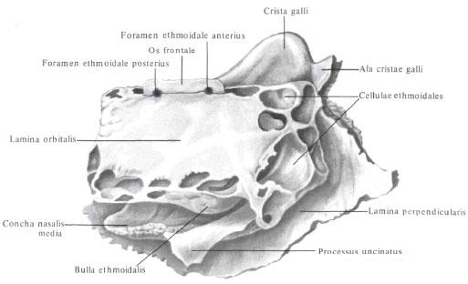
The trellis plate , lamina cribrosa, is the upper wall of the nasal cavity, is located horizontally in the trellis of the frontal bone , forming a front-latticed suture, sutura frontoethmoidalis. It is perforated by 30-40 small holes, foramina fibrosae, through which the nerves pass (fibers of the olfactory nerves) and vessels.
The perpendicular plate, lamina perpendicularis, is divided into two parts: a smaller upper plate lying above the trellis plate and a large lower one located under this plate. The upper part forms a cock's crest, crista galli, and is directed into the cavity of the skull (a crescent is attached to the crest of the cerebral cortex - the dura of the dura mater).
The border of the front edge of the cockscomb on each side is a non-permanent formation - the wing of the cock's crest, ala cristae galli. Both processes separate the blind hole from the back and the top, foramen cecum, the frontal bone. The lower part of the perpendicular plate of an irregular quadrangular form, directed vertically downwards, into the nasal cavity, and forms the anteroposterior part of the osseous septum. Above it is adjacent to the forehead of the frontal bone, in front to the nasal bones, behind - to the wedge ridge, from below - to the vomer , and from the front and from below - to the cartilaginous part of the septum of the nose. Often there is a deviation of all or part of the perpendicular plate to the side.

Grate labyrinth, labyrinthus ethmoidalis, - pair formation, located on both sides of the perpendicular plate, abuts the lower surface of the trellis plate. It consists of numerous air-bearing lattice cells, cellulae ethmoidales, communicating both with each other and with a series of holes with a nasal cavity. Latticed cells lined with a mucosa, which is a direct continuation of the nasal mucosa.
The cells located in front open in the middle nasal passage, the middle and posterior ones communicate with the superior nasal passage.
The lateral wall is a thin smooth orbital plate, lamina orbitalis, which forms the major part of the inner wall of the orbit. The plate connects at the top with the frontal bone, forming the frontal-latticed seam, sutura fronto-ethmoidalis, at the bottom - with the maxilla - the lattico-maxillary suture, sutura ethmoidomaxillaris, and with the orbital process of the palatine bone - the lumbar-suture seam, sutura palato-ethmoidalis, in front - with a tear bone - a tear -latticed seam and behind - with a sphenoid bone - a wedge-latticed suture, sutura spheno-ethmoidalis. Two small grooves run along the upper edge of the labyrinth: the anterior and posterior latticed grooves, which, with the same fissures of the frontal bone, form the tubules opening by the anterior and posterior trellised holes, foramina ethmoidales anterius and posterius (through which the same vessels and nerves pass through them).
The medial wall of the labyrinth is a rough, furrowed plate that forms most of the lateral wall of the nasal cavity. On its surface facing the perpendicular plate, there are two thin processes, slightly curved along the edges and wrapped outward: the upper - the upper nasal concha, the concha nasalis superior, and the lower one - the middle nasal concha, concha nasalis media. Sometimes a rudimentary process in the form of a thin bony scallop - the highest nasal conch, concha nasalis suprema - is located above the superior nasal shell. In the upper-posterior part of the medial wall of the labyrinth, between the upper and middle nasal shells, a space-upper nasal passage, meatus nasi superior-is formed. The slit under the middle nasal conch is the middle nasal passage, meatus nasi medius.
From the lower anterior surface of each labyrinth, anterior and posteriorly from the middle nasal cone, the hooked process bends posteriorly and downwards, processus uncinatus. On the whole skull it joins with the trellis process, processus ethmoidalis, inferior nasal concha.
Behind and up from the hook-shaped process is one of the largest cells, which looks like a bulge, - a trellis vesicle, bulla ethmoidalis.
Between the hook-shaped process from below and from the front and a large latticed bubble on the back and from above there is a gap - a crater funnel, infundibulum ethmoidale, the upper end of which communicates with the aperture of the sinus of the frontal bone. The posterior edge of the hook-shaped process and the lower surface of the large latticed vesicle form a semilunar cleft, hiatus semilunaris, through which the maxillary sinus communicates with the middle nasal passage.
You will be interested to read this:

Comments
When commenting on, remember that the content and tone of your message can hurt the feelings of real people, show respect and tolerance to your interlocutors even if you do not share their opinion, your behavior in the conditions of freedom of expression and anonymity provided by the Internet, changes Not only virtual, but also the real world. All comments are hidden from the index, spam is controlled.