Upper jaw
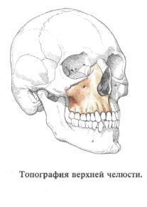
 Upper jaw , maxilla, paired, located in the upper front of the facial skull . It refers to the number of airborne bones, since it contains an extensive cavity lined with a mucous membrane, the maxillary sinus , sinus maxillaris.
In the bone, the body and the four processes are distinguished.
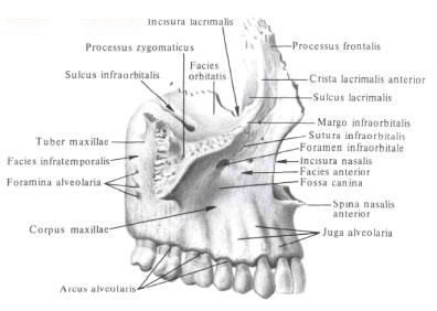
The body of the upper jaw , corpus maxillae, has four surfaces: the orbital, anterior, nasal and transverse.
Distinguish the following bone processes: frontal, malar, alveolar and palatine.
The facial surface, the facies orbitalis, is smooth, has the shape of a triangle, somewhat inclined anteriorly, outward and downward, forms the lower wall of the vagina, orbita.
The medial edge of it joins ahead with the tear bone , forming a tear -maxillary suture, posteriorly from the tear bone - with the orbital plate of the latticed bone in the latticotomycin seam and then backwards - with the orbital process of the palatine bone in the maxillofacial suture.


The anterior margin of the orbital surface is smooth and forms a free infraorbital margin, margo infraorbitalis. Being the lower part of the orbital margin of the orbit, margo orbitalis. Outside, it is serrated and passes into the zygomatic process. Medially the infraorbital margin forms a bend upward, sharpens and passes into the frontal process, along which the longitudinal anterior tear ridge stretches, crista lacrimalis anterior. At the place of transition to the frontal process, the inner edge of the orbital surface forms a lacrimal incision, incisura lacrimalis. Which, together with the teardrop cortex of the lacrimal bone, limits the upper opening of the nasolacrimal canal.
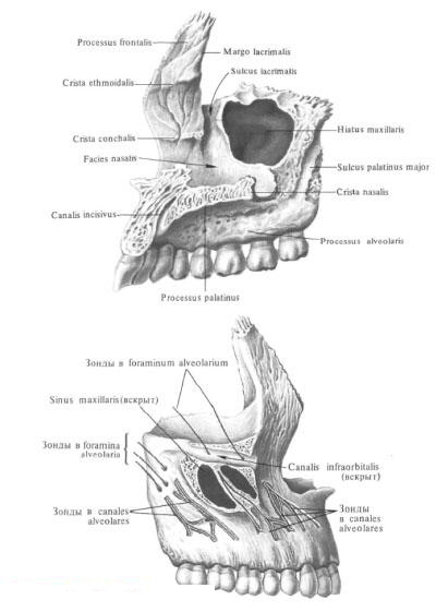
The posterior edge of the ophthalmic surface, together with the lower edge of the orbital surface of the large wings of the sphenoid bone parallel to it, forms the lower orbital fissure, fissura orbitalis inferior. In the middle part of the lower wall of the slit there is a groove - infraorbital furrow, sulcus infraorbitalis, which, going anteriorly, becomes deeper and gradually passes into the infraorbital canal, canalis infraorbitalis (in the furrow and to the palate the infraorbital nerve, artery and veins). The canal describes an arc and opens on the front surface of the body of the upper jaw. In the lower wall of the canal, there are many small holes of the dental tubules - the so-called alveolar orifices, foramina alveolaria; Through them the nerves pass to the group of anterior teeth of the upper jaw.
The subfamily surface, facies infratemporalis, faces the inframammary fossa, fossa infratemporalis, and the pterygopalatine fossa pterygopalatina, uneven, often convex, forms the tubercle of the maxilla, tuber maxillae. It distinguishes two or three small alveolar orifices leading to the alveolar canals, canales alveolares, through which the nerves pass to the back teeth of the upper jaw.
The anterior surface, fades anterior, is slightly curved. Below the infraorbital margin, a rather large infraorbital foramen opens on it, foramen infraorbitale, below which there is a small depression - a canine fossa, fossa canina (here the muscle that lifts the corner of the mouth, m. Levator anguli oris originates).
At the bottom, the anterior surface without a noticeable border passes into the anterior (buccal) surface of the alveolar processus, processus alveolaris, on which there is a series of convexities - alveolar elevations, juga alveolaria.
Inside and anteriorly, towards the nose, the anterior surface of the body of the upper jaw passes into the acute edge of the nasal tenderloin, incisura nasalis. At the bottom, the notch ends in the anterior nasal awn, spina nasalis anterior. The nasal incisions of both maxillary bones restrict the pear-shaped aperture, apertura piriformis, leading to the nasal cavity.
Nasal surface, facies nasalis, upper jaw more complex. In the upper-posterior corner there is an opening - maxillary cleft, hiatus maxillaris, leading to the maxillary sinus. Behind the crevices, a rough nasal surface forms a seam with a perpendicular plate of the palatine bone. Here on the nasal surface of the upper jaw vertically passes a large palatal sulcus, sulcus palatinus major. It is one of the walls of the large palatal canal, canalis palatinus major. Anterior to the maxillary fissure is a tear sulcus, sulcus lacrimalis, bounded in front by the posterior margin of the frontal process. To the tear sulcus there is a tearing bone at the top, below - a tear duct of the inferior shell. In this case, the lacrimal furrow closes into the nasolacrimal canal, canalis nasolacrimalis. Even more anterior to the nasal surface is a horizontal protrusion - shell ridge, crista conchalis. To which the lower nasal shell is attached.
From the upper edge of the nasal surface, at the place of its transition into the anterior, the frontal process, processus frontalis, is straightened up. It has a medial (nasal) and lateral (facial) surface. Lateral surface anterior lacrimal crista, crista lacrimalis anterior, divides into two sections - anterior and posterior. The posterior part downwards passes into the lacrimal furrow, sulcus lacrimalis. The border of it from the inside serves as a tear edge, margo lacrimalis. To which is adjoined a lacrimal bone, forming with it a tear-maxillary suture, sutura lacrimo-maxillaris. On the medial surface from front to back passes a trellis ridge, crista ethmoidalis. The upper margin of the frontal process is notched and joins with the nose of the frontal bone, forming the fronto-maxillary suture, sutura frontomaxillaris. The anterior edge of the frontal process is connected to the nasal bone in the naso-maxillary suture, sutura nasomaxillaris.
The spinal process, processus zygomaticus, departs from the outer upper corner of the body. The rough end of the zygomatic process and the zygomatic bone, os zygomaticum, form the maxillary maxillary suture, sutura zygomaticomaxillaris.
The palatine processus processus palatinus is a horizontally located bone plate that extends from the lower edge of the nasal surface of the body of the upper jaw and together with the horizontal plate of the palatine bone forms a bone septum between the nasal cavity and the oral cavity. Inner rough edges of the palatine processes, both maxillary bones join together to form the median palatine suture, the sutura palatina mediana. To the right and left of the seam there is a longitudinal palatal shaft, torus palatinus.
The posterior margin of the palatine process touches the anterior edge of the horizontal part of the palatine bone, forming with it a transverse palatal seam, sutura palatina transversa. The upper surface of the palatine processes is smooth and slightly concave. The lower surface is rough, near its posterior end there are two palatine fissures, sulci palatini, which are separated from each other by small palatine awns, spinae palatinae (in the furrows lie the vessels and nerves). The right and left palatine processes at the anterior margin form an oval-shaped incisal fossa, fossa incisiva. At the bottom of the fossa there are incisal holes, foramina incisiva (two of them), which opens the incisal canal, canalis incisivus. Terminating also with incisive holes on the nasal surface of the palatine processes. The canal can be located on one of the processes, in this case on the opposite process there is a incisive groove. The area of the incisal fossa from the palatine processes sometimes separates the incisal suture, sutura incisiva; In such cases a bone bone is formed, os incisivum.
Alveolar processus, processus alveolaris, the development of which is associated with the development of the teeth, departs from the lower edge of the body of the upper jaw and describes an arc directed by convexity forward and outward. The lower surface of this region is the alveolar arc, arcus alveolaris. On it there are lunochki - dental alveoli, alveoli dentales, in which there are roots of teeth - 8 on each side. The alveoli are separated from each other by interalveolar septa, septa interalveolaria. Some of the alveoli in turn are divided by inter-root septa, septa interradicularia, into smaller cells according to the number of tooth roots.
The anterior surface of the alveolar process, corresponding to the five anterior alveoli, has longitudinal alveolar elevations, juga alveolaria. Part of the alveolar process with the alveoli of the two anterior incisors represents a separate incisive bone in the embryo, os incisivum, which early merges with the alveolar process of the upper jaw. Both alveolar processes join and form an intermaxillary suture, sutura intermaxillaris.
You will be interested to read this:


Comments
When commenting on, remember that the content and tone of your message can hurt the feelings of real people, show respect and tolerance to your interlocutors even if you do not share their opinion, your behavior in the conditions of freedom of expression and anonymity provided by the Internet, changes Not only virtual, but also the real world. All comments are hidden from the index, spam is controlled.