


Temporal bone
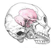
The temporal bone , os temporale, the parenteral, participates in the formation of the base of the skull and the lateral wall of its arch. In it lies the organ of hearing and balance. It articulates with the lower jaw and is the support of the chewing apparatus.
On the external surface of the bone there is an external auditory opening, porus acusticus externus, around which are located three parts of the temporal bone; On top - scaly part, inside and behind - rocky part, or pyramid, in front and below - drum part.
The scaly part, pars squamosa, has the shape of a plate and is located almost in the sagittal direction. The external temporal surface, facies temporalis, of the scaly part is slightly rough and slightly convex. In the posterior part it passes in the vertical direction the furrow of the middle temporal artery, sulcus arteriae temporalis mediae (imprint of the same artery).
In the posterior part of the scaly part there passes an arcuate line that extends into the lower temporal line, linea temporalis inferior, of the parietal bone .
From the scaly part, higher and somewhat anterior to the external auditory aperture, the zygomatic process extends horizontally, processus zygomaticus. It is as it were a continuation of the supra-succulent crista, crista supramastoidea, located horizontally along the lower edge of the outer surface of the scaly part. Beginning with a wide root, the malar spine then narrows. It has an inner and outer surface and two edges - a longer upper and lower, a shorter one. The anterior end of the zygomatic process is notched. The occipital process of the temporal bone and the temporal process, temporalis processus, the malar bone are connected by means of the temporal-zygomatic suture, sutura temporozygomatica forming the zygomatic arch, arcus zygomaticus.
On the lower surface of the root of the zygomatic process is a transversely oval shape of the mandibular fossa, fossa mandibularis. The anterior half of the fossa, up to the stony-scaly slit, is the articular surface, fades articularis, temporomandibular joint. Ahead of the mandibular fossa restricts the articular tubercle, tuberculum articulare.

The external surface of the scaly part participates in the formation of the temporal fossa,
Fossa temporalis (here begins the bundles of the temporal muscle , m. Temporalis).
Internal cerebral surface, facies cerebralis, slightly concave. It has finger-like indentations, impressiones digitatae, as well as arterial furrow, sulcus arteriosus (in it lies the middle meningeal artery, a. Meningea media).
The scaly part of the temporal bone has two free edges - wedge-shaped and parietal.
The anterior cuneate margin, margo sphenoidalis, broad, dentate, connects with the scaly margin of the large wing of the sphenoid bone and forms a wedge-scaly seam, sutura sphenosquamosa.
Upper-posterior parietal margin, margo parietalis, pointed, longer than the previous one, connected to the scaly edge of the parietal bone.
The pyramid (stony part), pars petrosa, the temporal bone consists of the posterolateral and anterior medial divisions.
The posterolateral part of the stony part of the temporal bone is the mastoid processus mastoideus, which is located posteriorly from the external auditory aperture. On it distinguish the outer and inner surfaces. The outer surface is convex, rough and is the place of attachment of muscles. The mastoid process turns into a conical protuberance, which is well probed through the skin,
On the inside, the process is restricted by a deep mastoid fillet, incisura mastoidea (it originates in the posterior abdomen of the digastric muscle, venter posterior M. digastrici). Parallel to the incision and somewhat posteriorly is the furrow of the occipital artery, the sulcus arteriae occipitalis (the imprint of the adherence of the same-named artery).
On the inner, medullary, surface of the mastoid process passes a wide S-shaped sigmoid sulcus, sulcus sinus sigmoidei, which passes at the top into the eponymous furrow of the parietal bone and further into the groove of the transverse sinus of the occipital bone (sinus transversa occurs in it). The furrow of the sigmoid sinus continues as the eponymous furrow of the occipital bone.
Behind the border of the mastoid process is a serrated occipital margin, margo occipitalis, which, connecting with the mastoid margin of the occipital bone, forms the occipital mastoid suture, sutura occipitomastoidea. At the middle of the seam length or in the occipital margin there is a mastoidal foramen mastoideum (sometimes several), which is the site of mastoid veins, vv. Emissariae mastoidea, connecting the subcutaneous veins of the head with the sigmoid venous sinus, as well as the mastoid branch of the occipital artery, ramus mastoideus a. Occipitalis.
Above the mastoid process is bounded by the parietal margin, which forms a parietal notch on the border with the same edge of the scaly part of the temporal bone, incisura parietalis; It includes the mastoid angle of the parietal bone, forming a parieto-mastoid suture, sutura parietomastoidea.
At the point of transition of the external surface of the mastoid process to the external surface of the scaly part, it is possible to notice remnants of the scaly-mastoid suture, sutura squamosomastoidea, which is well pronounced on the skull of children.
On the mastoid process, the bony airway cavities - mastoid cells, cellulae mastoideae - are located inside it. These cells separate one another from the bony mastoid walls, paries mastoideus. The permanent cavity is the mastoid cave, antrum mastoideum, in the central part of the appendage; It opens the mastoid cells, it connects to the tympanic cavity, cavitas tympanica. The mucous cells and the mastoid cave are lined with a mucous membrane.
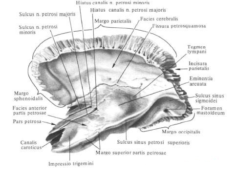
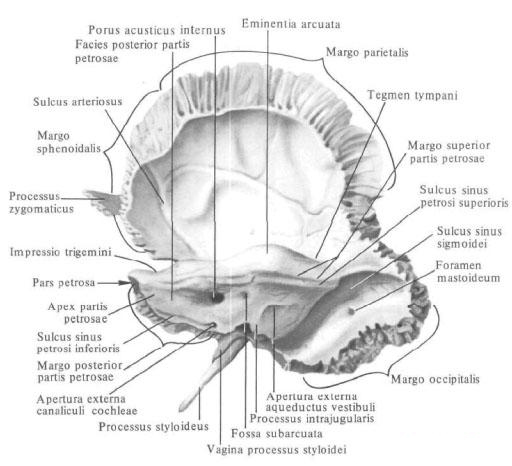
The anterior median part of the stony part lies inwardly from the scaly part and mastoid process. It has the form of a triangular pyramid, the long axis of which is directed from the outside and from behind and forward and medially. The base of the rocky part is turned to the outside and back; The apex of the pyramid, apex partis petrosae, is directed towards the inside and anteriorly.
In the rocky part, three surfaces are distinguished: the front, back and bottom, and three edges: the upper, the rear and the front.
The front surface of the pyramid, the facies anterior partis petrosae, is smooth and wide, facing the cavity of the skull, is directed obliquely from the top down and forward and passes into the medullary surface of the scaly part. From the latter it is sometimes separated by a stony-scaly slit, fissura petrosquamosa. Almost on the middle of the front surface there is an arcuate elevation, eminentia arcuata, which is formed by the underlying semicircular channel of the labyrinth lying beneath it. Between the elevation and the stony-scaly slit there is a small area - the roof of the tympanum, tegmen tympani, under which there is a drum cavity, cavum tympani. On the front surface, near the tip of the rocky part, there is a slight trigeminal impression, impressio trigemini (the junction of the trigeminal node, ganglion trigeminale).
Lateral to the depression is the cleft of the canal of the large stony nerve, hiatus canalis n. Petrosi majoris, from which a narrow fissure of a large stony nerve is medially guided, sulcus n. Petrosi majoris. Anterior and somewhat lateral from this opening is a small cleft in the channel of a small stony nerve, hiatus canalis n. Petrosi minoris, from which the groove of the small stony nerve is directed, sulcus n. Petrosi minoris.
The posterior surface of the pyramid, facies posterior partis petrosae, as well as the anterior one, faces the cranial cavity, but is directed upward and posteriorly, where it passes into the mastoid process. Almost in the middle of it there is a circular inner auditory aperture, porus acusticus internus, which leads to the internal auditory meatus, meatus acusticus internus (in it passes the facial, intermediate, pre-cochlear nerves, nn. Facialis, intermedius, vestibulocochlearis, and, artery And vein of the labyrinth, a. Et v. Labirinthi). A little higher and lateral from the inner auditory aperture there is a well-pronounced fossa subarcuata in newborns, of a small depth, fossa subarcuata (it includes the outgrowth of the hard shell of the brain). More laterally lies the slit-shaped outer aperture of the aqueduct of the vestibule, apertura externa aqueductus vestibuli, opening into the aqueduct vestibuli, aqueductus vestibuli. Through the aperture from the cavity of the inner ear emerges endolymphatic duct.
The lower surface of the pyramid, facies inferior partis petrosae, rough and uneven, forms part of the lower surface of the base of the skull. On it there is a round or oval jugular fossa, fossa jugularis (the place of adherence of the upper bulb of the internal jugular vein).
You will be interested to read this:

Comments
When commenting on, remember that the content and tone of your message can hurt the feelings of real people, show respect and tolerance to your interlocutors even if you do not share their opinion, your behavior in the conditions of freedom of expression and anonymity provided by the Internet, changes Not only virtual, but also the real world. All comments are hidden from the index, spam is controlled.