


Sky
The palate is the upper wall of the mouth proper. It is divided into a hard and soft sky. 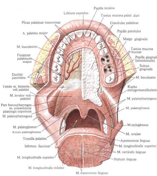
The front part of the sky is a solid sky , palatum durum, has a bone base - the bone skies , palatum osseum, which is formed by the palatine processes of the upper jaws and horizontal plates of the palatine bones. The back of the sky is the soft palate, palatum molle, mainly formed by muscles, aponeurosis and mucosa, in which the palatine glands are located.
The mucous membrane closely adjacent to the hard palate, smooth, passes from the front and from the sides to the gum, behind - to the soft palate, to its uvula palatina, and the arms of the sky. In the middle of the mucous membrane of the sky there is a narrow whitish strip - the seam of the palate, the raphe palati, near the medial incisors, there is a small fold - the incisive papilla, papilla incisva, which corresponds to the incisal canal, canalis incisivus.
From the seam in the transverse direction several (or one) weakly expressed transverse palatine folds, plicae palatinae transversae. In the seam area, the mucous membrane of the palate is thinner than at the edges. Between her and the periosteum is a thin layer of mucous membranes of the palatine glandulae glandulae palatinae. Forming two oblong clusters, they perform a space between the bone skies and alveolar processes.
The layer of glands of the hard palate thickens in the direction to the back and passes without a noticeable boundary into the layer of glands of the soft palate.
The soft palate , palatum molle, is formed mainly by muscles. It distinguishes the front horizontal part, which is the continuation of the hard palate, and the back part, which goes obliquely back and down. The soft palate is also called the palatine curtain, velum palatinum. Together with the root of the tongue, it restricts the isthmus of the pharynx. The palatine curtain is covered with a mucous membrane that fuses with a well-developed palatine aponeurosis, aponeurosis palatina, the place of attachment, the muscles of the soft palate. The soft palate in the middle is stretched in a small conical form of the palatine uvula palatina; On its front surface you can see the continuation of the seam of the sky.
On each side the palatal curtain passes into two arms. One - anterior - the tongue of the tongue, amis palatoglossus, - goes to the root of the tongue , the other - the posterior one - passes into the mucous membrane of the sidewall wall of the pharynx - the palatine pharyngeal arch, arcus palatopharyngeus. From above, as a result of the connection of the back surface of the tongue-and-tongue arch and the front surface of the pharyngeal arch, a semilunar fold is formed, plica semilunaris, bounding above the supramundary fossa, fossa supratonsillaris.
Between the palatal arms, the soft sky and the root of the tongue is the space through which the oral cavity communicates with the pharyngeal cavity - the isthmus of the pharynx, isthmus faucium, and the anterior rounded edge of it in the clinic is called the yawn, fauces.
A thin triangular fold, plica triangularis, of the mucosa partially covering the inner surface of the palatine tonsil extends from the back surface of the tongue-lingual arch. Narrow at the top, it is attached with its wide base to the lateral edge of the root of the tongue . Between its posterior edge and the tongue-and-tongue arch in front, a triangular amygdala fossa is formed at the back, a fossa tonsillaris, at the bottom of which is the palatine tonsilla, tonsilla palatina, which fills the entire fossa in adults.
Innervation: nn. Palatini majores et minores, incisivi.
Blood supply: aa. Palatina descendens, palatina ascendens; V. Palatina externa, plexus pterygoideus, plexus pharyngeus.
Palatine tonsils , tonsilla palatina, - paired bean-shaped formation. Tonsils are located on each side between the lingual-lingual and the hypoglossal arch in the amygdala fossa. Outdoors, the tonsil has a fibrous lining - amygdala capsule, capsula tonsillaris, and borders on the buccal part m. Constrictor pharingis superior. Its internal surface is uneven, with numerous round or oval amygdala dimples, fossulae tonsillares, corresponding to amygdala crypts, criptae tonsillares. The latter are depressions of the epithelial lining, lie in the substance of the palatine tonsil. Numerous lymphatic nodules, noduli lymphatici, are embedded in the walls of the pits and crypts.
In a normal state, the amygdala does not go beyond the fovea and there is free space above it - a supramaximal fossa, fossa supratonsillaris.
Innervation: nn. Palatini, n. Nasopalatinus, plexus palatinus.
Blood supply: a. Palatina ascendens (a. Facialis), a. Palatina descendens (a. Maxillaris), r. Tonsillaris a. Facialis. Venous blood from the sky is sent to v. Facialis. The lymph flows off into the nodi lymphatici submandibulares and submentales.
Muscles of the sky and throat.
1. Muscle straining the palatine, m. Tensor veli palatini, flat, triangular, located between the medial pterygoid muscle and the muscle lifting the palatal curtain. With its broad base, the muscle begins from the scaphoid fossa, fossa scaphoidea, the sphenoid bone, the membranous plate of the cartilaginous part of the auditory tube and the edge of its osseous furrow, reaching the spine of the sphenoid bone. Going downwards, it passes into a narrow tendon, which, circling the furrow of the pterygoid hook of the pterygoid process and the mucous bag on it, is then scattered by a broad bundle of tendon fibers in the aponeurosis of the soft palate. Some bundles are attached to the posterior edge of the horizontal plate of the palatine bone, partially intertwining with bunches of the same-named muscle on the opposite side.
Function: stretches the front section of the soft palate and the pharyngeal part of the auditory tube.
Innervation: n. Tensoris veli palatini.
2. The muscle lifting the palatal curtain, m. Levator veli palatini, flat, situated medially and posteriorly from the previous one. It starts from the lower surface of the stony part of the temporal bone, anterior to the outer opening of the canal channel, and from the cartilaginous part of the auditory tube, from the side of the lower medial surface.
The bundles are directed downward, inwards, forward and, expanding, enter the soft sky, interlacing with the beams of the same-named muscle of the opposite side. Part of the bundles is attached to the middle section of the sky aponeurosis.
Function: raises the soft palate, narrows the pharyngeal opening of the auditory tube.
3. Muscles of the tongue, mm. Uvulae, are two muscle bundles that converge to the midline of the tongue. The gradual decrease in the number of muscle beams causes its conical shape. Muscles originate from the posterior nasal awn of the hard palate, spina nasalis posterior, from the palatine aponeurosis and are directed to the median line, weave into the mucous membrane of the tongue. Most of the muscle bundles attached to the palatine aponeurosis reach the middle line, as a result of which the middle part is thickened and is called the seam of the sky.
Function; Shorten the tongue, lifting it.
4. The lingual-lingual muscle, m. Palatoglossus, narrow, flat, lying in the eponymous bow. The muscle begins from the lateral edge of the root of the tongue , forming, as it were, the extension of its transverse muscle bundles, and, rising to the top, ends in the aponeurosis of the soft palate.
Function: Narrows the pharynx, bringing the front arches together with the root of the tongue ,
5. Neo-pharyngeal muscle, m. Palato-phatyngeus, flat, triangular, most of it lies in the eponymous bow. The muscle begins with a wide base in the region of the posterior wall of the throat part of the pharynx and from the plate of the thyroid cartilage. Muscular bundles are directed to the middle of the sky and upward and enter from the sides into the thickness of the soft palate, where they are weaved into the palatine aponeurosis. Some of the bundles are attached to the pterygoid hook of the pterygoid process, and a part to the lower edge of the medial plate of the cartilage of the auditory tube and forms the tubal-pharyngeal muscle, m. Salpingopharyngeus. Function: brings the palate to the pharyngeal arch and pulls up the lower part of the pharynx and larynx.
Innervation: all four muscles are plexus pharyngeus.
Blood supply: all muscles are aa. Palatinae (a. Facialis, a. Maxillaris).
You will be interested to read this:

Comments
Commenting on, remember that the content and tone of your message can hurt the feelings of real people, show respect and tolerance to your interlocutors even if you do not share their opinion, your behavior in the conditions of freedom of expression and anonymity provided by the Internet, changes Not only virtual, but also the real world. All comments are hidden from the index, spam is controlled.