


Colon, structure, functions
The colon , colon, in its position, as it were, fringes the loops of the small intestine located in the middle of the lower floor of the abdominal cavity. Ascending colon is on the right, transverse - on top, descending - on the left, sigmoid - on the left and partly from below.
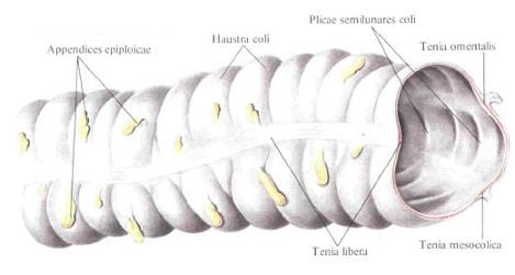
Ascending colon , colon ascendens, begins from the place of confluence into the cecum of the ileum, being a continuation of the blind. It is separated from the caecum by two grooves that correspond to the bridles of the ileocecal valve. Its posterior, devoid of peritoneum surface adjoins the posterior abdominal wall, occupying the extreme lateral position to the right. It starts slightly below the iliac crest, rising vertically, first in front of the square muscle of the waist, further ahead of the right kidney and reaches the lower surface of the right lobe of the liver; Here it bends to the left and the ventral (forward) and passes into the transverse colon. The bend is called the right flexure of the colon, flexura coli dextra, and compared to the left flexure of the colon, flexura coli sinistra, is usually more flat. Due to the fact that the right bend is directed not only in the frontal but also in the sagittal plane, the initial part of the transverse colon lies superficial or in front of the ascending one (this also applies to the left bend). The length of the ascending colon reaches 20 cm, but its position and length are quite variable: often with a high position of the caecum the ascending colon has a length of 12 cm or even less. The mites on the ascending colon are arranged in the following sequence: on the front surface - a free tape, tenia libera, on the posterolateral - the glandular tape, tenia omentalis, and on the posterior medial - mesenteric tape, tenia mesocolica.
The transverse colon , colon transversum, begins in the right subcostal area at the X level of the costal cartilage from the right flexure of the colon, goes a few oblique directions from right to left and upwards to the left subcostal area. Here, at level IX of the costal cartilage or the eighth intercostal space, it passes to the descending colon in the left flexure of the colon. The left portion of the transverse colon lies superficial (ventral) to the descending colon. The middle part of the transverse colon crosses the epigastric region, forming a downwardly directed bend (sagging), so that the ascending and descending colon together with the transverse colon resemble the letter M. The length of the transverse colon reaches 50 cm. This is the longest section of the large intestine. It is located intraperitoneally and has its own mesentery, mesocolon transversum, starting on the back wall of the abdomen from the parietal peritoneum.
To the anterior surface of the transverse colon along the extension of the posterolateral gland ribbon, tenia omentalis, a gastric-ligament ligament is attached, lig. Gastrocolicum, is part of a large omentum, an omentum majus that covers all parts of the small intestine. As a result of this arrangement, the transverse colon, concealed by an epiploon in the front, is not visible or only shines when the abdominal cavity is opened. If you unscrew the gland together with the transverse colon facing up to its rear surface, you can see its posterior (dorsal) surface with a free ribbon, tenia libera, and a mesenteric transperum mesentery.
The left flexure of the colon, flexura coli sinistra, is located in the left hypochondrium region, much higher and deeper (dorsally) than the right one, immediately below the lower pole of the spleen. The left end of the transverse colon forms an acute angle with the initial section of the descending colon, the apex of which is fixed by a leaflet of the peritoneum descending from the diaphragm (lig. Phrenicocolicum).
The descending colon , colon descendens, is located on the back wall of the abdomen, occupying here the extreme left position at the side wall. Begins at the top of the left bend and descends the back wall of the abdomen; Its posterior, devoid of peritoneal surface lies in front of the lateral region of the left kidney and the square muscle of the waist and reaches the level of the left iliac crest; Here goes into the next section of the colon - the sigmoid colon. The descending colon is lateral to the midline of the abdomen than the ascending one. Its length is more than ascending, and reaches 22-23 cm. The diameter of the intestine of the previous sections of the colon and at the level of the transition to the sigmoid colon is 4 cm. The amount of the caustic and their depth decrease; The location of the muscle bands, the position of the peritoneum and glandular processes is the same as on the ascending colon.
The sigmoid colon , colon sigmoideum, is located in the left ileal fossa. It starts from the top and laterally at the level of the posterior edge of the iliac crest. Having formed two loops, one of them, proximal, located on the iliac muscle, convex downward, and the other, distal, located on the large lumbar muscle, is turned upward, the sigmoid colon goes right (medially) and downward, bends through the border line and Enters the cavity of the small pelvis, where at level III of the sacral vertebra passes into the rectum. The length of the sigmoid colon is on the average 54-55 cm, it is subject to significant individual fluctuations (from 15 to 67 cm); Its diameter is about 4 cm. The sigmoid colon is located intraperitoneally and has a mesentery.
The structure of the walls of the blind and colon has its own characteristics. Completely of three layers - the peritoneum, the muscular and the mucous membranes - only those parts of the large intestine that are located intraperitoneally, namely: the blind, transverse colon, sigmoid colon and upper third of the rectum; The ascending colon and the descending colon (in some cases, the caecum) have a peritoneal cover on three sides: lateral, anterior and medial.
The site of the posterior wall of the ascending colonic and descending colon is 2-3 cm wide and devoid of serosa; Mesenteric parts of the large intestine - transverse and sigmoid colon - have a narrow, peritoneum-free band along the attachment line of the mesentery. In the locations of the furrows on the colon, the serous membrane follows the wall behind the indentation.
In rare cases, the lower parts of the ascending and descending colon can be covered with a serous membrane from all sides and even form mesentery.
The muscular membrane, tunica muscularis, throughout the colon forms two layers - the outer longitudinal layer, stratum longitudinale, and the inner circular, circular layer, stratum circulare. The longitudinal layer over most of the stretch is assembled into strips. The appendix has a continuous two-layered muscular cover, which is weaker than in other parts.
The mucosa, tunica mucosa, consists of an epithelial cover with underlying basal membrane, an intrinsic connective tissue layer and a muscular plate of the mucous membrane, lamina muscularis mucosae, under which lies the submucosa, tela submucosa.
The epithelium of the mucosa consists of cylindrical cells with a large number of goblets. The mucosa of the colon contains intestinal glands, glandulae intestinales, but is devoid of villi. Throughout the mucous membrane there are single lymphatic follicles, folliculi lymphatici solitarii. Corresponding to the location of the transverse grooves, the mucosa forms semilunar folds of the colon, plicae semilunares coli.
At the site of the ileum in the thick - ileocecal opening, ostium ileocecale, there are two permanent folds of the intestinal wall, mainly from the circular muscle layer. They form an ileocecal valve, valva ileocecalis. The edges of the orifices are fused and continue in the form of a bridle of the ileocecal valve, frenulum valvae ileocecalis, located on the border of the blind and ascending colon. At the base of the shutter, the circular muscle layer is more developed, forming a kind of pulp.
The mucous membrane of the appendix is characterized by an abundance of lymphoid tissue, forming an almost continuous layer in the form of group lymphatic follicles of the appendix, folliculi lymphatici aggregati appendicis vermiformis.
You will be interested to read this:

Comments
Commenting on, remember that the content and tone of your message can hurt the feelings of real people, show respect and tolerance to your interlocutors even if you do not share their opinion, your behavior in the conditions of freedom of expression and anonymity provided by the Internet, changes Not only virtual, but also the real world. All comments are hidden from the index, spam is controlled.