| Start of section
Production, amateur Radio amateurs Aircraft model, rocket-model Useful, entertaining |
Stealth Master
Electronics Physics Technologies Inventions |
Secrets of the cosmos
Secrets of the Earth Secrets of the Ocean Tricks Map of section |
  |
| Use of the site materials is allowed subject to the link (for websites - hyperlinks) | |||
Navigation: => |
Home / Patent catalog / Catalog section / Back / |
|
INVENTION
Patent of the Russian Federation RU2154409
![]()
METHOD OF RESEARCH OF ENGINE FUNCTION OF THE ESOPHAGUS AND PROBES FOR ITS IMPLEMENTATION
The name of the inventor: M.Abakumov ; Pinchuk TP; Volkov, S.V .; Popova TS; Vasilyev VA; Azarov Ya.B.
The name of the patent holder: Moscow City Research Institute of Emergency Care. N.V. Sklifosovsky
Address for correspondence: 129010, Moscow, B. Sukharevskaya pl., 3, Research Institute of First Aid to them. N.V. Sklifosovsky, the patent group
Date of commencement of the patent: 1999.02.15
The invention relates to medicine, in particular to gastroenterology, and can be used to diagnose esophagus diseases accompanied by a violation of its motor function. The technical result is an extension of the diagnostic capabilities of the method by performing a complex assessment of the esophagus motor function, including a local measurement of intraluminal pressure and measuring the electrical impedance of the entire esophageal wall at various levels. To do this, a two-channel probe is introduced into the proximal part of the stomach, one of the channels of which is filled with liquid and is intended for measuring intraluminal pressure, the other for measuring the impedance of the esophageal wall. The proximal end of the probe is connected to a recording device from which the recorded values are transferred to the computer. The first 4 - 5 minutes register these values with the static position of the probe, then the probe is slowly pulled up to the upper esophageal sphincter, making a mark on the curve about the position of the probe in the esophagus. After extraction of the probe, the results obtained are analyzed. A differential-digestive feature is a violation of the synchronism of changes in intracavitary pressure and electrical impedance during their registration.
DESCRIPTION OF THE INVENTION
The invention relates to medicine, in particular to gastroenterology, and can be used to diagnose esophagus diseases accompanied by a violation of its motor function.
The wide prevalence of diseases of the esophagus and cardia in recent years has contributed to the isolation of esophagology into a separate area of gastroenterology (Tamulevichyute DI, Vitenas AM "Diseases of the esophagus and cardia", Moscow, Medicine, 1986, p.3. The important role belongs to the functional methods of research, in particular, the determination of the motor function of the esophagus and cardia (Vasilenko V.X., Suvorova TA, Grebenev AL "Akhalasia cardia." Moscow, Medicine, 1976, p. Disorders of motor function of the esophagus themselves cause a number of painful symptoms, in addition, they are an important pathogenetic link in diseases such as hernia of the esophagus of the diaphragm reflux-esophagitis, with acute damage to the esophagus (Tamulevichute DI, Vitenas A M., "Diseases of the esophagus and cardia." Moscow, Medicine, p.58-60) Further progress in the treatment of these diseases is associated with the improvement of diagnostic methods ("Wege zur Diagnose - Entscheidungsprozesse in der Medizin". Urban. Munchen, 1992, s. 152-157).
There is a known method for recording the motor function of the esophagus and cardia by measuring the intraepithelial pressure simultaneously at different levels of the esophagus (Vantsyan EN, Fedorova OD, Chissov VI, Tarutin VN Ezofagomanometric studies with functional obstruction of the esophagus. Functional obstruction of the digestive tract, M. 1967. pp. 149-158.) It consists in inserting into the body of the stomach a probe consisting of 2-5 connected open catheters with a diameter of 1 mm each, the distal ends of which are located at a distance of 2- 3 cm apart. The proximal end of the probe connects to pressure sensors, the number of which corresponds to the number of catheters in the probe. In turn, each pressure sensor is connected to the channel of the recording device, which allows recording pressure changes in real time in the form of a graph. After inserting the probe through the catheters, the liquid is passed under low pressure to create a hydraulic column. The indicator of the pressure in the catheter after stopping the fluid supply corresponds to the pressure created by the column of fluid at the open end of the catheter located at this point and is equal to the pressure in the lumen of the esophagus at its given point.
The disadvantage of this method is that: 1 / the pressure registered at a given point of the body is an indirect characteristic of the motor function of the muscles of the local part of the esophagus, since it represents the resultant value determined by the contraction of the muscles of this part of the esophagus and the distant muscles as well as the muscles of the stomach and pharynx . 2 / the described method makes it possible to obtain indicators of intraluminal pressure only at separate points of the organ corresponding to the localization of the distal ends of the catheters and does not allow us to judge the state of the motor function of the site of the organ between two points.
The object of the invention is to expand the diagnostic capabilities of the method by performing a complex assessment of the esophagus motor function in acute injuries and functional diseases of the esophagus, which includes both a local measurement of intraluminal pressure and the measurement of the electrical impedance of the entire esophageal wall at various levels.
The physiological basis for the possibility of recording electrical impedance from the esophageal wall is that the esophagus is a hollow organ with tightly closed walls and does not contain air and liquid (Saks FF, Baytinger VF, Medvedev MA, Ryzhov A. I. "Functional morphology of the esophagus", Moscow, Medicine, 1987, p. 86). The probe electrodes are in tight contact with its walls. The current fed to the current-setting electrodes passes through the organ wall, and the detected impedance corresponds to the impedance of the esophageal wall, the main component of which is the muscle envelope.
To solve the task in the method of examining the motor function of the esophagus by measuring intraluminal pressure in various parts of the esophagus, the electrical impedance of the esophagus wall is measured simultaneously with the help of probe electrodes inserted into the lumen of the esophagus, and then the results obtained at each monitoring stage are compared.
The probe for examining the motor function of the esophagus contains two channels, one of which has a lateral opening at the proximal end and is intended for fluid injection, the other channel contains electrodes with conductors for recording the impedance from the esophageal wall, with current-setting electrodes located at the ends of the probe, The entire length of the recording electrodes is located.
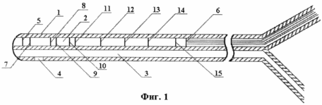
In Fig. 1 shows the general view of the probe.
The probe 1 comprises two channels: a channel 3 with a diameter of 1 mm, with a lateral opening 4 at the proximal end for introducing fluid, the channel 2 contains electrodes with conductors, with current-setting electrodes 5, 6 located 25 cm apart at the edges of the probe, and recording Electrodes 7-15 are located between the current-setting electrodes along the entire length of the probe. The electrodes 7, 15 are removed from the current-setting electrodes at a distance of 0.5 cm. The electrodes 8.9 and 10, 11 are separated from each other by a distance of 1 cm and 9.10 by a distance of 2 cm. Between the pairs of electrodes 7-8, 11 -12, 12-13, 13-14, 14-15 - a distance of 4 cm. Above the location of the electrodes on the probe there are labels that allow to determine the localization of each electrode with respect to the front teeth (incisors) or to the wings of the nose. Channel 3 with the adapter is connected to the pressure sensor. The electrodes of channel 2 are connected to the electrical impedance recording device connected to the computer.
The method is carried out as follows. In the horizontal position of the patient on the back through the oral cavity or nasal passage, the probe is inserted into the stomach in such a way that the electrodes 5, 6 are located in the proximal part of the stomach, the electrodes 8, 9, 10, 11 in the lower esophageal sphincter, electrodes 12, 13, 14 - in the body of the esophagus, electrodes 15-16 in the region of the superior esophageal sphincter. The localization of these parts of the esophagus with respect to the incisors in each particular patient is determined by the endoscope label with the endoscopic examination performed the day before. After insertion of the probe, the channel 3 is connected to a pressure sensor, which in turn is connected to a water supply device (dropper), the channel 3 is filled with a liquid (physiological solution). Then, the probe electrodes through the conductors of channel 2 are connected to a device measuring the electrical impedance at an alternating current frequency of 50-100 kHz. All signals are recorded at an analog-to-digital conversion frequency of at least 5 Hz for each signal with a resolution of at least 12 bits. Digital representation of the signals is input to the computer for real-time monitoring (monitoring), saving and subsequent processing.
Monitoring of the study is carried out in two stages. At the first stage, the impedances and pressure in the static position of the probe are recorded for 4-5 minutes. Periodically, the patient is asked to swallow saliva. At the second stage, the probe is gradually raised for 4-5 minutes, noting the level of the probe's location in centimeters from the wings of the nose or from the incisors on the curve. In the process of pulling the patient's probe and asking to do swallowing movements.
After extraction of the probe, the results obtained are analyzed.
The indices of intraluminal pressure were determined according to a conventional method. The impedance curves at rest were used to determine the rate of peristaltic wave passage during swallowing movements, the appearance of sharp jumps or high-frequency contractions, the relative level of impedance in the proximal part of the stomach and various parts of the esophagus. The parameters obtained by pulling the probe made it possible to evaluate changes in intraluminal pressure at a certain level of the esophagus in parallel with changes in the impedance of the corresponding part of the organ sequentially along the entire esophagus. In this case, the peaks in the pressure and impedance curves were determined: a differential diagnostic feature is a violation of the synchronism of changes in intracavitary pressure and electrical impedance during their registration, a reaction to swallowing movements and the presence of spontaneous contractions.
The proposed method was used to examine 10 healthy and 37 patients with acute injuries and chronic diseases of the esophagus: chemical burn of the esophagus, cardiopathism, reflux esophagitis, spasm of the superior esophageal sphincter, hiatal hernia. The analysis of the results obtained made it possible to distinguish the following variants of motor dysfunction of the esophagus.
Normally, there was no spontaneous, non-absorbent contraction on the impedance curves and pressure, and when swallowed, a synchronous increase in impedance and pressure was recorded.
Periodic short-term decrease in the impedance in the lower esophagus allowed to diagnose the transfer of gastric contents into the esophagus - gastroesophageal reflux.
The increased excitability of the muscles of the esophagus was characterized by: 1 / the appearance of multiple peaks of contractions on the impedance curve at rest in the absence of changes in the pressure curve ("muscle flicker"), 2 / the appearance of multiple peaks of contractions on the impedance curve in response to a sip that corresponded to only one peak of increase Intraluminal pressure.
Reduced excitability of the muscles of the esophagus (inhibition of motor function) was characterized by a low-amplitude increase in intraluminal pressure in the absence of rise peaks on the impedance curve (i.e., intraluminal pressure is increased only by contraction of the pharyngeal muscles).
A comparison of the indices of the impedance and the intraluminal pressure makes it possible to differentiate the cases of increase in the intra-esophageal pressure due to the gastric component. Thus, if there are peaks in the pressure increase in the pressure curve in the absence of reduction peaks on the impedance curve, the latter is due to the spread of gastric tension on the esophagus (spasmic contractions of the stomach in patients with gastric stasis, chemical burn of the stomach).
Clinical examples:
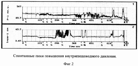
1. Patient B., 42 years old. Has arrived with the diagnosis: a chemical burn of an esophagus of 3 degrees, a chemical burn of a stomach of 3 degrees by an electrolyte, on EGDS an esophagus from 25 sm from cutters with a circular touch of fibrin. Cardia is not completely closed. In the body of the stomach and subcardia, a circular fibrin deposit. Local treatment with laser irradiation has been started. Esophagoimpedanosomonometry has been performed (Figure 2). As can be seen, along with the peaks of the reduction in the impedance curve and the pressure that appear on the pharynx, there are spontaneous peaks in the increase in the intra-esophageal pressure, which indicates spasmodic contractions of the stomach.
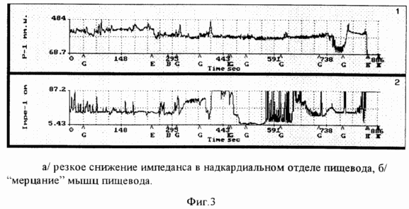
2. Patient A., 57 years old. Has arrived with the diagnosis: a chemical burn of an esophagus 2 degrees with an acetic essence. The irradiation of the mucosa of the esophagus with a helium-neon laser was performed - 10 sessions. At a control examination visually (EGDS), cardia deficiency, erosive reflux esophagitis is determined. Ezofagoimpedan-somanometry was performed (Figure 3). The impedance curve shows a sharp decrease in the endocardial section of the esophagus. There is an increased excitability of the muscles of the esophagus, when in response to a sip a series of peaks appears on the impedance curve, which corresponds to only one peak of increase in the intra-esophageal pressure. There are also extra-glossy contractions on the impedance curve with no change in the intra-esophageal pressure ("flicker").
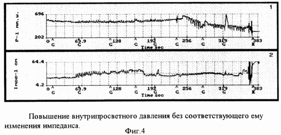
3. Patient A., 71 years old. Has arrived with complaints about pains in epigastric area. With EGDS revealed catarrhal reflux-esophagitis, insufficiency of cardia. Ezofagoimpedanosomanometry was performed. When comparing the impedance and pressure curves (Figure 4), it can be seen that in the distal esophagus after swallowing saliva there is an insignificant increase in the intra-esophageal pressure and there is no corresponding impedance that indicates inhibition of the contractile function of the esophagus muscles and an increase in intraluminal pressure only due to the pharyngeal component .
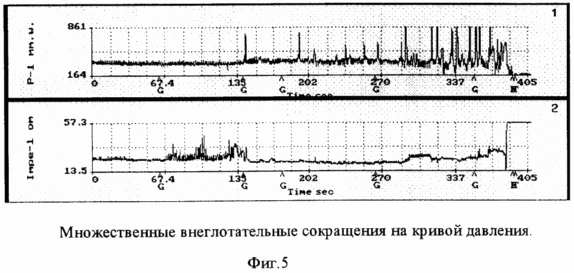
4. Patient A., 35 years old. Has arrived with the diagnosis: a chemical burn of an esophagus 3 degrees, a chemical burn of a stomach of 2 degrees an unknown cauterizing substance. On the EGDS, the esophagus mucosa is all over with erosive-ulcerative changes. Cardia is not completely closed. The mucous membrane of the stomach is hyperemic, edematic, with focal patches of fibrin. Ezofagoimpedanosomanometry was performed. On the pressure curve (Fig. 5), multiple swallowing contractions are noted, while the impedance curve did not change. In this observation, the increase in intra-esophageal pressure is associated with an increase in intragastric pressure.
Thus, the proposed method allows in detail to differentiate the motor function of the esophagus and cardia in its functional diseases and injuries.
CLAIM
1. A method of examining the motor function of the esophagus by measuring intraluminal pressure in various parts of the esophagus, characterized in that the electrical impedance of the esophagus wall is measured simultaneously with the help of probe electrodes inserted into the lumen of the esophagus, and then the results of measurements are compared at each monitoring stage.
2. A probe for examining the motor function of the esophagus, comprising a channel with a lateral opening at the distal end for introducing a fluid, characterized in that it further comprises a channel with electrodes for recording the impedance of the esophageal wall, the current-setting electrodes being located at the ends of the probe, and between them throughout The recording electrodes are located long.
print version
Date of publication 29.03.2007gg
![]()

Comments
When commenting on, remember that the content and tone of your message can hurt the feelings of real people, show respect and tolerance to your interlocutors even if you do not share their opinion, your behavior in the conditions of freedom of expression and anonymity provided by the Internet, changes Not only virtual, but also the real world. All comments are hidden from the index, spam is controlled.