
| Start of section
Production, amateur Radio amateurs Aircraft model, rocket-model Useful, entertaining |
Stealth Master
Electronics Physics Technologies Inventions |
Secrets of the cosmos
Secrets of the Earth Secrets of the Ocean Tricks Map of section |
  |
| Use of the site materials is allowed subject to the link (for websites - hyperlinks) | |||
Navigation: => |
Home / Patent catalog / Catalog section / Back / |
|
INVENTION
Patent of the Russian Federation RU2265404
![]()
METHOD OF PROPHYLAXIS OF INCONSISTENCY OF CULTIVA OF CEREBRAL EXTRACT
The name of the inventor: Bilichenko V.B. (RU); Nazarenko PM (RU); Mezentsev A.I. (RU); AN Danilov (RU); Nagapetyants A.G.
The name of the patent holder: Bilichenko Vyacheslav Borisovich
Address for correspondence: 305048, Kursk, Druzhby Ave., 15, apt.191, VB Bielichenko
Date of commencement of the patent: 2000.10.20
The invention relates to medicine, gastroenterology, can be used in the surgical treatment of destructive forms of acute appendicitis. Band and cut off the appendix at the base. The cult of the appendix is covered with a mobilized fat suspension of the ileocecal angle. The suspension is sewn to the dome of the cecum by 3-4 P-shaped serous-muscular sutures. The method allows to prevent chronic intestinal obstruction, reflux-ileitis.
DESCRIPTION OF THE INVENTION
The invention relates to medicine, namely to gastroenterology, and can be used for surgical treatment of destructive forms of acute appendicitis complicated by tiflite.
The closest to the claimed solution is the method of surgical treatment of destructive forms of acute appendicitis complicated by tiflite, in which the stalk of the appendix is covered by the stump of the vermiform appendage (AM Antonov, Yu.B.Volkov, NA Yaitsky, KM. Chernov, MV Grigorieva, VN Grinenko "The inconsistency of the stump of the appendix after appendectomy", Herald of Surgery, 1999, №2).
However, the use of this method in the surgical treatment of destructive forms of acute appendicitis complicated by tiflite can lead to deformation of the ileocecal angle and cause the development of complications such as chronic intestinal obstruction, reflux ileitis.
The object of the invention is to prevent the development of chronic intestinal obstruction, reflux-ileitis.
The task is accomplished by the fact that the stump of the appendix is covered by the mobilized fat suspension of the ileocecal angle, which is sewn to the dome of the cecum by 3-4 P-shaped serous-muscular sutures.
 |
 |
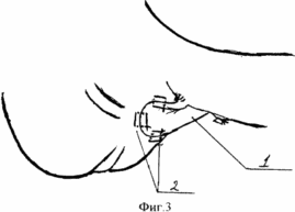 | |
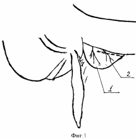
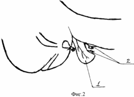
Figures 1, 2, 3 are schematic diagrams for explaining the invention.
The method is carried out as follows. The laparotomy is performed and a dome of the cecum with a vermiform appendage is excreted into the wound. Mesentery of the appendix is stitched, bandaged and cut off. After this, the base of the appendix is ligated at the dome of the cecum. Then, in cases of pronounced tiflitis, which is manifested by thickening and swelling of the cecal wall around the base of the appendix, proceed in fact to perform the proposed method, since it is typical to immerse the stump of the appendix into the pouch, and then the Z-shaped suture is impossible due to the eruption of the filaments. In the wound, the fat suspension of the ileocecal angle is removed. (1) and mobilize its top. For this, the outer vessels (artery and vein) are stitched and bandaged (2) in FIG. 1, after which the apex of the gut is cut off from the gut (1, 2. FIG. 2). Then the suspensions (1) of Fig. 3 are laid on the stump of the appendix and sutured to the dome of the cecum by 3-4 P-shaped serous-muscular sutures (2. Fig. 3), thus covering the stump of the appendix. The operation is terminated by removal of the exudate, drainage of the abdominal cavity with glove-tubular drainage, administration of antibiotics and layer-by-layer suturing of the wound.
The invention is illustrated by the following example
Patient M., 24 years old, case history No. 1047, entered the surgical department of the hospital with a clinic of acute destructive appendicitis on 20.09.99.
At admission, he complained of aching pain in the right iliac-inguinal region, nausea, a single vomiting, an increase in body temperature, a general weakness.
He is sick during the day, when pains appeared in the epigastric region for the first time, which then in 6 hours shifted to the right iliac-inguinal region. He applied for medical help one day after the illness. The emergency ambulance was delivered to the hospital.
At admission, the condition of the patient is of medium severity, the skin is of normal color, warm. From the side of lymphatic, osteoarticular, muscular systems without features. Body temperature is 37.5 ° С. The frequency of respiratory movements is 20 per minute, respiration is vesicular, there is no wheezing. Heart sounds are clear, rhythmic, no noise, heart rate 94 beats per minute, blood pressure 120/80 mm Hg. Art. The tongue is clean, dryish. The abdomen is of the correct configuration, the anterior abdominal wall is limitedly involved in the act of breathing due to lagging of the right abdominal parts. With superficial palpation, muscle tension of the anterior abdominal wall and soreness in the right iliac-inguinal region are noted. At a deep palpation the expressed morbidity in the right ileal-groin area is defined. Symptoms of Voskresensky, Sitkovsky, Bartome-Mikhelson, Shchetkin-Blumberg are positive. The liver, the gallbladder is not palpated. When auscultation - peristalsis somewhat weakened in the right abdomen. Urination free, painless. The gases leave, there was no chair.
The general analysis of the blood: hemoglobin 143 g / l, erythrocytes 4.1 × 10 12 / l, leukocytes 12.0 × 10 9 / l, stab 10; Segmented 72, monocytes 2, and lymphocytes 16.
General analysis of urine: color straw-yellow, specific gravity 1020, pH acidic, protein 0,066 g / l, microscopy of sediment: leukocytes 4-6 in sp., Erythrocytes 1-2 in sp., Cylinders hyaline 1-2 In the sp., Oxalates a small amount.
Ultrasound data: no signs of pathology are seen from the kidneys, gall bladder; A vermiform appendix with dimensions of 8 × 2.5 cm and a wall thickness of 4 mm is visualized.
Based on the clinic, laboratory and instrumental examination data, a diagnosis of acute destructive appendicitis and indications for surgery was established.
20.09.99 1 hour after the patient has received the operation. Under intravenous anesthesia, an oblique incision through the McBurney-Volkovich-Dyakonov in the right ileal region revealed the abdominal cavity. At the same time up to 20 ml of turbid effusion without odor. The effusion is drained. A dome of the cecum with a vermiform appendage was excreted into the wound. The latter is sharply enlarged in size - 8 × 2.5 cm, strained, hyperemic, covered with fibrinous overlays. The wall of the caecum around the base of the appendix is thickened, edematous, hyperemic. Mesentery of the appendix is stitched, bandaged and cut off. After this, the base of the appendix is tied up at the dome of the cecum. Then, taking into account the phenomena of tiflitis, manifested by thickening and edema of the cecal wall around the base of the appendix, it was decided to abandon the typical immersion of the stump of the appendix into the pouch and then the Z-shaped suture due to probable eruption of the filaments. The terminal site of the ileum with the fat suspension of the ileocecal angle was removed into the wound and its apex mobilized. For this, the marginal vessels (artery and vein) are stitched, the upper part of the intestine is cut off. After this, the suspensions are laid on the stump of the appendix and sutured to the dome of the cecum with three U-shaped serous-muscular sutures, thus the stalk of the appendix is covered with a fat suspension. The operation was completed by draining the abdominal cavity with glove-tubular drainage, adding 0.5 g of kanamycin to 20.0 ml of physiological saline and draping the wound with layer-by-layer drainage. The postoperative course is smooth. Drainage is removed on the 3rd day, the stitches are removed on the 7th day. Recovery.
FIG. 1 depicts the fatty suspension of the ileocecal angle (1), the terminal vessels of the suspension (2).
2 depicts the mobilized fatty suspension of the ileocecal angle (1), the marginal vessels (artery and vein) are stitched and ligated (2). The appendix is cut off.
Figure 3 shows the final form of the operation - the fatty pendant (1) is sutured with 3 U-shaped serous-muscular sutures (2) to the dome of the cecum, covering the stump of the appendix.
CLAIM
A method for preventing the inconsistency of the stump of the appendix, including bandaging and cutting off the appendix at the base, characterized by the fact that the stump of the appendix is covered by a mobilized fat suspension of the ileocecal angle, which is sutured to the dome of the cecum by 3-4 P-shaped serous-muscular sutures.
print version
Date of publication 29.03.2007gg
![]()

Comments
When commenting on, remember that the content and tone of your message can hurt the feelings of real people, show respect and tolerance to your interlocutors even if you do not share their opinion, your behavior in the conditions of freedom of expression and anonymity provided by the Internet, changes Not only virtual, but also the real world. All comments are hidden from the index, spam is controlled.