| Start of section
Production, amateur Radio amateurs Aircraft model, rocket-model Useful, entertaining |
Stealth Master
Electronics Physics Technologies Inventions |
Secrets of the cosmos
Secrets of the Earth Secrets of the Ocean Tricks Map of section |
|
| Use of the site materials is allowed subject to the link (for websites - hyperlinks) | |||
Navigation: => |
Home / Patent catalog / Catalog section / Back / |
|
INVENTION
Patent of the Russian Federation RU2198603
![]()
METHOD OF TREATMENT OF GASTROESOPHAGEAL REFLUX DISEASE BY RECOVERY OF VALVE GUBAREV'S FUNCTION OVER DIAPHRAGMA
The name of the inventor: Zalewski AA
The name of the patent holder: Krasnoyarsk State Medical Academy
Address for correspondence: 660022, г.Красноярск, ул. Partizan Zheleznyak, 1, Medical Academy, Patent Department
The effective date of the patent: 2001.02.21
The invention relates to medicine, gastroenterology, can be used in the treatment of gastroesophageal reflux disease. Mobilize the left lobe of the liver. Dissect the esophagus-diaphragmatic ligament or membranous diaphragm at the anterior edge of the ring of the esophageal opening of the diaphragm. Mobilize the hernial part of the stomach and 5.0 cm of the esophagus. Separate the diaphragm from the pericardium by 3.0 cm in width and anterior to the posterior line of their adhesion. Sew the anterior edge of the esophageal-gastric junction with the pericardial surface of the diaphragm 3 cm anterior to its esophageal opening. Sew the edges of the diaphragm section. The method restores the function of the Gubarev valve over the diaphragm.
DESCRIPTION OF THE INVENTION
The invention relates to medicine and can be used for the treatment and prevention of gastroesophageal reflux disease (GERD).
The views of modern surgeons on the factors of GERD pathogenesis, as objects of surgical correction, are not the same. Many refer to them a hernia of the esophageal opening of the diaphragm (GVPD), an enlarged angle of the Gysus and the esophageal aperture of the diaphragm, a weak lower esophageal sphincter (NPC), a non-hermetically esophagogastrial valve (Gubareva), and subject them to surgical correction. Often in the foreground is the desire to at any cost to eliminate the HVAC. For this, a high mobilization or plastic lengthening of the esophagus is performed by Collis and a reduction of the cardia under the diaphragm. With even greater consistency, the pressure of the NPS is increased by wrapping the distal esophagus with a cuff from the bottom of the stomach (the Nissen, Toupe, and Dora operations and their modification) and restoring the acute angle of the Giss by cross-linking the stomach bottom with the esophagus to normalize the Gubarev valve function [1, 2, 4 , 10, 13].
We believe that the restoration of the angle of the Hyis by the above method is not sufficient to restore the function of the valve, because Due to the long stay in the stretched state, the lower rings of the NPS lose the ability to contract. Therefore, the valve remains leaking. The force data of the NPC does not play a role in this case [6, 8, 9, 11].
Concerning the repositioning of the cardia under the diaphragm, with a shortened esophagus of the 2nd degree, the opinions diverge. Clinic BV Petrovsky and her followers prefer to preserve the mediastinal dislocation of the cardia, in order to avoid tension and slippage of the esophagus from the cuff of Nissen (telescope phenomenon) after surgery and relapse of GERD [3, 5, 10].
A large number of modifications of "cuff" operations means dissatisfaction with their results and complications. The most constant and frequent ones are postoperative dysphagia, atony and bloating, impossibility of eructation and vomiting, rupture of the cuff, relapse of heartburn [4, 5, 13, 14].
Having studied the role of the Gubarev valve in the pathogenesis of reflux esophagitis [6], we came to a somewhat different conclusion than the other authors: "To stop the GER, it is sufficient to restore the antireflux function of the Gubarev valve."
We realized our plan, only not in the traditional way. First of all, we focused on correcting only the key factors of the pathogenesis of GERD and minimizing the operating injury. Therefore, the degree of esophagus shortening, its mobility, the strength of the NPS, we considered in patients as variants of the individual norm, not subject to surgical correction.
Objective of the invention. To develop a method for treating gastroesophageal reflux disease by restoring the function of the Gubarev valve over the diaphragm.
The task is solved due to the upper middle laparotomy, mobilization of the left lobe of the liver, dissection of the esophagus-diaphragmatic ligament or membrane diaphragm at the anterior end of the esophageal ring of the diaphragm, mobilization of the hernial part of the stomach and 5 cm of the esophagus, separation of the diaphragm from the pericardium by 3.0 cm in width And anterior to the posterior line of their fusion, stitching the anterior edge of the esophageal-gastric junction with the pericardial surface of the diaphragm 3.0 cm anterior to its esophageal opening, stitching the edges of the diaphragm cut.
The procedure of the operation. Perform upper-median laparotomy. Mobilize and divert the left lobe of the liver. The finger is attempted to pass between the anterior edge of the esophageal ring of the diaphragm (CPD) and the anterior wall of the stomach. If the finger passes into the hernial sac, then lower the stomach downwards under the diaphragm, dissect and excise the hernial sac leaving the edge (KGM) in the esophagus. Mobilize up to 5.0 cm of esophagus and take on the holder.
 |
 |
If the EFFECT and the front wall of the stomach are firmly fused, then we are dealing with a congenital short esophagus. To dissect these fuses is dangerous because of the possibility of damage to the trunk of the left vagus nerve and the wall of the stomach. Therefore, an oval incision of the diaphragm is made at the anterior margin of the CPD. Through the incision, the chest stomach is mobilized and about 5.0 cm of the esophagus. Take them on the holder. (Fig. 1). The anterior edge of the CPD is withdrawn posteriorly. A finger or a prepuce gauze stump on a curved clip exfoliates the pericardium from the diaphragm by 3.0 cm in width and anterior to the posterior line of their adhesion. Under the PZP line, the front wall of the stomach is stitched with three U-shaped ligatures, their ends tied and taken in pairs on the clamps. The double ends of the ligatures are threaded into a large arcuate needle and through the CPD or its incision from the top downwards, through the periocardial part of the diaphragm separated at a distance of 3.0 cm anterior to its margin, while maintaining parallelism with the sagittal line. From the side of the abdominal cavity, the ends of the ligatures are stretched and moved by the PZP to the line of their stitching through the diaphragm. The double ends of the side ligatures are connected to the individual ends of the middle ligature (FIG. 1). After that, the anterior walls of the cardiac part of the stomach and esophagus form an acute angle (similar to the angle of the Hyis) directed anteriorly and bounded in front and below by the diaphragm, and in front and from above by the pericardium of the posterior circle of the heart (Fig. Then remove the holder and sew the edges of the diaphragm cut.
The posterior walls of the cardiac section of the stomach and esophagus repeat the course of their anterior walls. Helps them in this NPC.
Essential longitudinal tension of the esophagus does not occur, t. The place of fixing the PZP to the diaphragm is much higher than the CPD.
 |
Elements forming the Gubarev valve and the principle of its antireflux function. When the NPC is in a tone, its posterior wall is at the anterior wall, 3.0 cm anterior to the CPD. PZHP is closed and hermetically sealed from below by the anterior wall of the cardiac part of the stomach, fixed to the diaphragm and enveloping it from front to back (fixed valve flap). The posterior wall of the cardiac part of the stomach lies on the anterior and further seals the PCI (Fig. 2). During the swallowing act, the NPS relaxes, but does not expand arbitrarily. Its walls push apart the food passing through the esophagus. The posterior wall of the cardia and the cardiac part of the stomach (flap) departs posteriorly and upward. The food passes unhindered into the esophagus (FIG. 3). The NPS again comes in a tone, closes the cardia, brings its rear wall to the front and "hides" the PZHP behind the fixed valve flap. The flap returns to the stationary one and seals the threshold of the PWR (Fig. 2). Cardia is not limited from the outside and is fixed to the diaphragm only by the anterior margin. Therefore, its lumen expands to the maximum possible, and any force of the NPS is sufficient to close the PZP and start behind the fixed valve flap. |
During physiological relaxation of NPS, not associated with food intake, the pressure in the esophagus remains low, and in the abdominal cavity - high. Therefore, the cardia remains asleep, the flap is fixed and the GER does not occur. This is confirmed by the 24-hour pH-metry.
5. Abdominal access of such operations is performed 5. Excellent results in all patients. Immediately after the operation, GERD symptoms disappeared. There were no signs of dysphagia in any patient. They should not be, because NPCs are not wrapped with a cuff.
An example from practice . Patient M. 61 years old, medical history 3686. Received in the 1st Surgical Department on 27.11.00 with the diagnosis: GERD of the 3rd stage, erosive-ulcerative reflux esophagitis. Concomitant diseases (aggravating) chronic stone cholecystitis in remission, hormone-dependent bronchial asthma, hypertensive disease, ischemic heart disease.
 |
 |
The indication for the operation was frequent exacerbations of GERD and concomitant exacerbations of bronchial asthma. Radiologic examination revealed a GAP, a second shortened esophagus of the 2nd degree, gastroesophageal reflux (Fig. 4). The main symptoms were: heartburn, chest pain, dysphagia, nocturnal regurgitation of gastric contents, attacks of bronchial asthma. After medical preoperative therapy on 31.11.2000, operations were performed: antireflux surgery according to this method, removal of the gallbladder with calculi. The postoperative period has passed with a moderate exacerbation of bronchial asthma. It took an increase in the dose of hormonal drugs within 5 days. There were no other complications. They were discharged on recovery for 21 days, from the date of admission to the hospital.
 |
 |
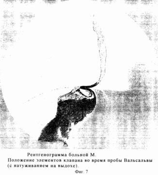
On radiographs performed 19 days after the operation, in the prone position on the abdomen with a raised left side, the moment of passage of barium through the valve zone during the act of swallowing the barium suspension (Figure 5) and the time of its completion are recorded, when the esophageal walls and valve flaps still retain Traces of barium contrast (Fig. 6). The flap returned to the fixed one and covered the PZP. Three minutes after the sip of the barium slurry, a Valsalva test was performed. There is no reflux of contrasting mass into the esophagus (Fig. 7).
Thus, the antireflux function of the Gubarev valve restored above the diaphragm reliably prevents the GER, without correcting the pressure of the NPS.
The proposed method for treating GERD is simple in execution. It does not provide for the reduction of cardia under the diaphragm and therefore is relatively non-traumatic. The effective function of the valve, regardless of the state of esophageal mobility and tone of the NPS, makes this method of treating GERD universal. It does not require preoperative dynamic studies of the esophagus and NPS, With them do not connect the key cause of the pathogenesis of GERD.
INFORMATION SOURCES
1) Alekseenko AV, Reva VB, Sokolov V.Yu. "Choice of a method of plasty with hernia of the esophageal opening of the diaphragm" - Surgery. - 2000. - 10. P. 12-14.
2) Galimov OV, Sakhautdinov VG, Senderovich EI, Fedorov SV "To the technique of fundoplication in the surgical treatment of reflux esophagitis." Vesti chir., 1997. T. - 156, 3, p. 47-48.
3) Kanshin NN, Chisov VI "Valvular gastroplication with a short esophagus 11-degree". - Surgery. - 1969. 12. - P. 55-58.
4) Kubyshkin VA And Fedorov VD, Kornyak BS, Azimov R.Kh. "The place of laparoscopic surgery in gastroesophageal reflux disease". - Surgery. - 1999. -. 11. - P. 4-7.
5) Oskretkov VI, Gankov VA "Results of surgical correction of cardia deficiency". Surgery, 1997, 8, 43-46.
6) Petrovsky BV, Kanshin NN, Nikolayev N.O. "Surgery of the diaphragm." Medicine. L. Otd., 1966, p. 175.
7) Shalimov AA, Sayenko VF, Shalimov SA "Surgery of the esophagus" - M .: Medicine. - 1975. - P. 116 and 109.
8) Sheptulin AA, Khromov VL, Sankina EA "Modern concept of pathogenesis, diagnosis and treatment of reflux esophagitis" Klin. honey. - 1995. - 6. - P. 11-14.
9) Efendiyev VM, Kasumov NA "Surgical correction of disorders of the cardiac closure function." Surgery - 1999. - 6 - P. 27-30.
10) Alien MS, Trastek VF, Deschamps C., Pairolero PC "Intrathoracic stomach. Presentation and results of operation" J. Thorac. Cardiovasc. Surg. - 1993 Feb. - Vol. 105. - N. 2. - P. 253-8.
11) Castell DO New York. - 1985. - P. 3-9.
12) Gastal OL, Hagen JA, Peters JH, Campos GM, Hashemi M., Thisen J., Bremner CG, DeMttster TR "Short esophagus: analisis of predictors and clinical implications" Arch. Surg. - 1999 Jun. - Vol. 134.-N. 6. - P. 633-6.
13) Kabat J., Pafko P. "Chrurgike osetreni refluxni nemoci jicnu pri kratkem jicnu" Rozhl. Chir. - 1994 Nov. - Vol. 73. - N. 7. - P. 345-7.
14) O Hanrahan T., Marples M., Bencewicz J. "Recurrent reflux and wrap disruption after Nissen fundoplication: Detection incidence and timing". G. Surg. - 1990, - vol. 77, No. 5 - P. 545-547.
15) Ramel S., Thor K., "The ersta procedure, a hemifundoplication for the treatment of gastroesophageal reflux disease" Ann. Chir. - 1995. - Vol. 84.-N. 2. - P. 145-9.
CLAIM
The method of treatment of gastroesophageal reflux disease by restoration of the function of the Gubarev's valve above the diaphragm, consisting of an upper median laparotomy, mobilization of the left lobe of the liver, dissection of the esophageal-diaphragmatic ligament or membrane diaphragm at the anterior end of the esophageal ring of the diaphragm, mobilization of the hernial part of the stomach and 5.0 cm of the esophagus, The separation of the diaphragm from the pericardium by 3.0 cm in width and anterior to the posterior line of their fusion, stitching the anterior edge of the esophagogastric junction with the pericardial surface of the diaphragm 3.0 cm anterior to its esophageal opening, stitching the edges of the diaphragm cut.
print version
Date of publication 29.03.2007gg


Comments
When commenting on, remember that the content and tone of your message can hurt the feelings of real people, show respect and tolerance to your interlocutors even if you do not share their opinion, your behavior in the conditions of freedom of expression and anonymity provided by the Internet, changes Not only virtual, but also the real world. All comments are hidden from the index, spam is controlled.