
| Start of section
Production, amateur Radio amateurs Aircraft model, rocket-model Useful, entertaining |
Stealth Master
Electronics Physics Technologies Inventions |
Secrets of the cosmos
Secrets of the Earth Secrets of the Ocean Tricks Map of section |
  |
| Use of the site materials is allowed subject to the link (for websites - hyperlinks) | |||
Navigation: => |
Home / Patent catalog / Catalog section / Back / |
|
INVENTION
Patent of the Russian Federation RU2200476
![]()
METHOD OF GASTRAP REACTION
The name of the inventor: Yu. P. Novomlinets; Zatolokin V.D .; Voinov S.A.
The name of the patent holder: Novomlinets Yuri Pavlovich
Address for correspondence: 305033, Kursk, K. Marx, 3, Meduuniversitet, Patent Department
Date of commencement of the patent: 2000.02.21
The invention relates to medicine, surgical gastroenterology, can be used to treat complicated forms of peptic ulcer of the stomach and duodenum. Resect two thirds of the stomach. In the region of the stomach, a small curvature is stitched with the ligature-holder. On a large curvature at 1.5 cm proximal to the pyloric sphincter, the terminal vessels are wall-banded. In this case, a large curvature of the stomach is mobilized to the level of the third distal branch of the left gastro-omental artery. Pristenochno mobilize a small curvature of the stomach proximal to the ligature-holder to the subcardial part of the stomach. The ligature-holder is pulled by a small curvature of the stomach to the right and upwards. From the initial point of mobilization of the suprapiloric section of the large curvature of the stomach, clamps are placed in the direction of the clamping ligature. Cross the stomach between the clamps so that the width of the distal and proximal stumps is the same. Apply an oblique suprapyloric gastrogastroanastomosis. The method allows to restore a normal passage of food through the gastrointestinal tract.
DESCRIPTION OF THE INVENTION
The invention relates to the field of medicine, namely surgical gastroenterology, and can be used to treat gastric ulcers complicated by bleeding, penetration, perforation, and duodenal ulcer.
A method of surgical treatment of peptic ulcer by performing a pylor-preserving resection of the stomach is proposed. Shalimovym. A.A. Shalimov, V. A. Saenko. Surgery of the digestive tract, 1987, 183. It consists in the mobilization and resection of 1/2 of the stomach from a level of 2 cm proximal to the pylorus. At the same level, the right gastric and right gastro-omental arteries intersect, the pyloric sphincter and the distal stump of the stomach are deprived of parasympathetic and sympathetic innervation. Pilor-preserving resection of the stomach according to the technique of A.A. Shalimova is one of the effective methods for preventing most post-resection syndromes, but the denervation of the pyloric sphincter and distal stump of the stomach that occurs during surgery often leads to motor-evacuation disorders in the early postoperative period.
Part of the task of restoring the normal passage of food through the gastrointestinal tract is solved by operating procedures aimed at preserving the nerves of Letarje and the main gastric circulation. These techniques suggest a seromyotomy of the entire small curvature of the stomach or an antrum flap and excision of the mucosa from these fragments. This is a long and time-consuming manipulation.
The method of resection of the stomach, proposed by S.S.Sh., is closest to solving the problem of ensuring the preservation of the innervation of the pyloroentral section and the distal stump of the stomach. Nikulshinym in 1986. Wedge. Surgery, 1988, 8, 72-73. The mobilization of a large curvature of the stomach from the suprapiloric section to the first branch of the left gastro-omental artery, producing the main trunk of the right gastric artery, is produced. Produce a wall-mounted mobilization of small curvature from the corner of the stomach to the esophagus, retaining the trunk of the left gastric artery and the nerves of Letarje. Proximal to the place of occurrence of Letharge's nerves in the wall of the stomach in the area of its angle with a scalpel dissect the serous and muscular membranes of the anterior and posterior walls of the stomach to the submucosa in parallel with a small curvature and deviate from it 1.0-1.5 cm to the level of distal mobilization and 2.0 Cm proximal to the pyloric sphincter. Then the serous-muscular flap is bluntly and sharply separated from the submucosa base and taken to the side. Supratrimonial part is crossed transversely by 2.0 cm proximal to the pylorus, and then the stomach is resected along the proximal mobilization boundary. Small curvature of the stomach and gastrogastroanastomosis are sewn with double-row stitches. The detached serous-muscular flap is sewn with separate nodular sutures from the stump of the stomach by a small curvature. Evaluation of long-term results of pylor-saving resection using SS technique. Nikulshina showed no dumping syndrome in all 24 of the examined patients, while 8 patients had a slowdown in the evacuation of contrast mass during X-ray examination and relapse of peptic ulcer after 2 years after surgery in one patient.
The received results testify to insufficient safety of function of nerves Letarzhe after mobilization of a serous-muscular flap according to SS technique. Nikulshina.
The aim of the invention is to increase the effectiveness of treatment of complicated forms of peptic ulcer and duodenal ulcer.
The task is achieved by the fact that pyloreconservation resection of 2/3 of the stomach is performed with preservation of the full parasympathetic and sympathetic innervation and blood supply of the pyloric sphincter and distal stump of the stomach by imposing an oblique suprapiloric gastrogastroanastomosis.
 |
 |
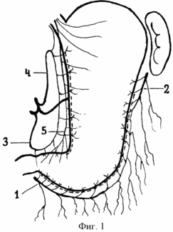
1 shows the boundaries of mobilization of the stomach in large and small curvatures, where 1 is the main trunk of the right gastro-omental artery, 2 is the main trunk of the left gastro-omental artery, 3 is the main trunk of the right gastric artery, 4 is the main trunk of the left gastric artery , 5 - Letarje nerve.
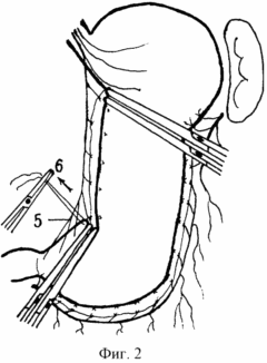
Fig. 2. The technique of imposing clamps on the mobilized fragment of the stomach is shown, where 5 is the Letarche nerve, 6 is the direction of traction of the ligature-holder, superimposed on the small curvature of the stomach proximal to the place where Leverage nerve enters the stomach.
THE METHOD IS PROVIDED BY THE FOLLOWING.
After the upper-median laparotomy, a revision of the stomach, ulcers, a study of the place where the Letraje nerve occur. Proximal, in the region of the stomach corner, a small curvature is stitched with the master ligature. Due to the large curvature 1.5 cm proximally to the pyloric sphincter, strictly pristenochno, banding only terminal vessels of the second order and retaining the main trunk of the right gastro-omental artery, mobilization of the large curvature of the stomach to the level of the third distal branch of the left gastro-omental artery. Proximal to the level of overlapping ligature-holder from the angle of the stomach to the subcardial part of the stomach is the post-wall mobilization of small curvature along the anterior and posterior surface of the stomach (Fig. 1). The ligature-holder small curvature of the stomach stretches to the right and up. From the initial point of mobilization of the suprapiloric section of the large curvature of the stomach towards the ligature-holder, rigid clamps are placed at the corner of the stomach and a stomach intersects them. At the level of the proximal border of the mobilization of the stomach, two rigid clamps are placed and the mobilized 2/3 of the stomach are cut off (Fig. 2). The mobilized stomach is located in the clamps so that the width of the distal and proximal stump is the same. The proximal and distal stump of the stomach easily converge. According to the conventional technique, an oblique suprapiloric gastrogastroanastomosis is imposed by two radial nodular sutures. Ends operation omentogastropeksiey. In the presence of a complicated duodenal ulcer, one of the variants of duodenoplasty is performed beforehand.
EXAMPLE OF EXPERIMENTAL IMPLEMENTATION.
In 10 mature mongrel dogs weighing from 8 to 15 kg, a pylor-saving resection of 2/3 of the stomach with oblique gastro-gastroanastomosis was performed while maintaining the blood supply and innervation of the gastroduodenal transition. The operations were performed under colicapsal anesthesia.
Dog B. 12 kg. The upper-median laparotomy was performed. A post-wall mobilization of the large curvature of the stomach was made from the level 1 cm proximal to the pylorus and to the third distal branch of the left gastro-omental artery. On the anterior and posterior surfaces of the stomach, a small curvature is mobilized from the corner of the stomach to the subcardial department. On the small curvature of the stomach at the level of the fourth branch of the Letarje nerve, a ligature-holder is applied. For her, the stomach wall was stretched to the right and upwards. The suprapiloric section of the stomach is expanded and clamped in an oblique direction from the large curvature 1 cm proximal to the pyloric sphincter to a small curvature in the region of the stomach angle. In parallel, the second clamp is applied. The second pair of clamps is applied to the body of the stomach at the level of the proximal mobilization boundary. The intersection of the stomach between the clamps in the distal and proximal part was made. The proximal and distal stomach stumps are close to each other. According to the common technique, two oblong seams of lavsan and catgut are overlaid with supra-pilor gastrostastroanastomosis. Anterior omega-gastropexy was performed. The carried out researches on experimental animals showed the technical possibility of performing pylorecaving resection of 2/3 of the stomach with oblique suprapilarological gastrogastroanastomosis and preservation of the full innervation and blood supply of the gastroduodenal transition. Morphological, X-ray, endoscopic examinations performed within 20-60 days showed healing of the stitches on the stomach by primary tension, revealed its good blood supply. There were no cases of insufficiency of sutures, stenosis, bleeding from the anastomosis line, development of acute pancreatitis, gross fusion of the walls of the stomach and abdominal wall. After 60 days after the operation, the motor-evacuation function of the pyloroduodenal transition was of a batch type for 2 h 30 min and did not differ from the function in healthy dogs. The phenomena of reflux gastritis were absent, positive dynamics of the weight of animals was noted.
EXAMPLE OF CLINICAL EXECUTION.
1. Patient Z., 38 years old. Case report 4851/543. I entered the clinic with a diagnosis of a callous giant ulcer of the upper third of the stomach. The peptic ulcer is ill for 3 years. It was treated out-patient. FGDS, fluoroscopy of the stomach and duodenum was performed. A chronic ulcer measuring 3.0 X 4.5 cm of the upper third of the stomach was detected. The preoperative preparation was carried out and the pylor-saving oblique resection of 2/3 of the stomach was performed. After laparotomy, a deformation and shortening of the small omentum was detected due to an inflammatory infiltrate in the upper third of the body of the stomach. A post-wall mobilization of the large curvature of the stomach was made from a level 1.5 cm above the pyloric sphincter to the third distal branch of the second order of the left gastro-omental artery. The level of penetration of three distal branches of the Letarje nerve was detected and a ligature-holder was applied to the stomach wall by 1 cm proximal to the small curvature. From her in the proximal direction to the subcardial part of the stomach along the front and back wall, his mobilization is carried out. Ligature-holder, imposed in the region of the angle of the stomach, stretched to the right and upward, lifting the wall of the stomach. On the suprapiloric stomach section in an oblique direction from the beginning of mobilization of large curvature, and in the direction proximal to the clamping ligature, rigid clamps are applied. Between the clamps the stomach is crossed. On the border of the proximal mobilization of the stomach and two rigid clamps are applied in such a way that the width of the formed proximal stump corresponds to the distal suprapiloric stump. Between the superimposed clamps the stomach is crossed in its upper third. For the purpose of hemostasis, the vessels of the submucosa of the proximal and distal stumps are sewn along the anterior and posterior surfaces. Two rows of nodal sutures lavsanom and vikril put gastrogastroanastomoz with preservation of the main blood supply of the stomach and three branches of the nerve Letharget, going to the pyloric sphincter and suprapiloric part of the antral part of the stomach. The anterior gastroenteropexy was performed and the small omentum was stitched to the stomach. The postoperative period proceeded without complications. Peristalsis of the intestine was restored by the end of 2 days, on the third day there was an independent chair.
In the control X-ray examination of the stomach and duodenum 2 weeks after the operation, small dimensions of the cylindrical configuration of the stomach stalk with the invaginated edges of oblique suprapiloric gastrogastroanastomosis were found, the pyloric sphincter was not deformed, a portion passage of the contrast mass into the duodenum was noted. Its bulb is not deformed, there is no duodenogastric reflux, the duration of evacuation of the contrast mass is 3 h 5 min. After 6 and 12 months, control EGF. There were no pathological changes in the resected stomach. At x-ray examination at 6 and 12 months, the gastrogastroanastomosis line is not determined. The motor-evacuator function of the pyloroduodenal transition corresponds to the normal parameters, is rhythmic, portioned, carried out within 2 hours. The patient performs the former work of the driver. Has added in weight of 7 kg, complaints are not present.
2. Patient P., 42 years old. History of the disease. Entered the clinic with a diagnosis of perforated callous ulcer bulb of the duodenum. There is no ulcerative anamnesis. Entered 1 hour after the perforation. Upon receipt, gas was detected under the right dome of the diaphragm. An emergency laparotomy was performed. Diffuse serous peritonitis. The abdominal cavity is sanitized. On the front wall of the bulb of the duodenum is a perforation with a diameter of 5 mm. Ulcerative infiltrate with a diameter of 30 mm. A rhomboid longitudinal excision of the infiltrate with partial excision of the pyloric sphincter was performed. Two rows of nodal sutures catgut and lavsan produced an enlarged anterior duodenoplasty. Mobilization of the large curvature of the stomach from the suprapiloric section to the third distal branch of the left gastro-omental artery was performed. In the region of small curvature of the stomach 1 cm proximal to the second distal branch of the Letarje nerve, a ligature-holder is applied. From the ligature in the proximal direction to the subcardial part of the stomach, a small curvature was mobilized along the anterior and posterior walls. A small curvature of the antral part of the stomach is stretched to the right and upwards for the ligature-holder. In an oblique direction on the suprapiloric department and the proximal part of the stomach, rigid clamps are placed and the mobilized part of the stomach is excised. Two lines of nodal sutures catgut and lavsan are overlaid with supra-pilor gastrostastroanastomosis. The abdominal cavity is drained by glove-tubular drainage in the hypochondrium and iliac regions. Postoperative course without features. On the third day there was an independent chair. On the 12th day I was discharged for outpatient care.
X-ray control 2 weeks after discharge showed a good function of the gastroduodenal transition, a portioned inflow of contrast into the duodenum during 2 hours and 50 minutes. Subsequent follow-up studies at 6 and 12 months showed a reduction in the time for evacuation of the contrast to 2 hours and 20 minutes. There are no signs of reflux gastritis, esophagitis. Has added in weight of 5 kg. He works on the previous work as a turner. He does not observe a diet.
According to the proposed technique, 10 patients underwent surgery. In 8 patients, the operation was combined with various variants of duodenoplasty. Given that the formation of gastrogastroanastomosis according to the technique of SS. Nikulshina and AA Shalimova the same type, we compared the results of the application of pylor-preserving resection in A.A. Shalimov and the methodology we proposed. In both groups, 10 cases were studied. The analysis of complications of the early and the nearest postoperative periods in both groups is presented in the table.
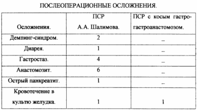 |
The obtained results show that the early and the nearest postoperative period was more favorable in patients after pyloreconservation resection of 2/3 of the stomach with oblique suprapiloric gastrogastroanastomosis. After pyloreconservation resection by the method of A.A. Shalimov more often develop complications associated with impaired patency of the anastomosis in the early postoperative period and motor-evacuation complications in the immediate postoperative period. The case of bleeding in the 1st day of the postoperative period in the stump of the stomach after pylor-saving resection with oblique gastro-gastroanastomosis did not require surgical intervention. |
Analyzing the literature data after the pylor-saving resection of the stomach, performed according to the method of SS Nikulshin, postoperative complications were assessed in 32 patients in terms of up to 10 years. Complaints about the feeling of stomach overflow after eating, pain, belching were noted by 15 patients, in 8 there was a delay in evacuation of the contrast mass from the stump of the stomach, in one patient there was a relapse of peptic ulcer of the stomach after 2 years. There were no signs of dumping syndrome. S.S. Nikulshin Surgery. 1993; 2: 14-16.
Thus, the proposed method of gastrectomy reduces the number of intra- and postoperative complications, reduces the likelihood of recurrence of peptic ulcer, excludes the development of dumping syndrome, reduces the number of motor-evacuation disorders of the gastrointestinal tract. There were no cases of gastrostasis or diarrhea, postoperative pancreatitis. The probability of postoperative bleeding in the stump of the stomach decreases, as the seam is not formed due to the small curvature of the resected stomach, the anastomosis is not impaired due to anastomosis, as the anastomosis is laid throughout the breadth of the lumen of the stomach stump, simplification and acceleration of the resection of the stomach.
CLAIM
The method of gastric resection for complicated forms of peptic ulcer of the stomach and duodenum, including resection of two thirds of the stomach, characterized by the fact that in the region of the stomach corner, small stiffness is stitched with a master ligature, a 1.5 cm proximal to the pyloric sphincter, Mobilizing a large curvature of the stomach to the level of the third distal branch of the left gastro-omentum artery, the mammary curvature of the stomach proximal to the ligature of the stomach until the subcardial part of the stomach is pristine, the small curvature of the stomach pulling to the right and upwards, from the initial point of mobilization of the suprapiloric section of the large curvature of the stomach Direction of the holder ligature, attach clamps and cross the stomach between them in such a way that the width of the distal and proximal stumps is the same, after which an oblique suprapiloric gastrogastroanastomosis is imposed.
print version
Date of publication 30.03.2007gg
![]()

Comments
When commenting on, remember that the content and tone of your message can hurt the feelings of real people, show respect and tolerance to your interlocutors even if you do not share their opinion, your behavior in the conditions of freedom of expression and anonymity provided by the Internet, changes Not only virtual, but also the real world. All comments are hidden from the index, spam is controlled.