Chewing muscles
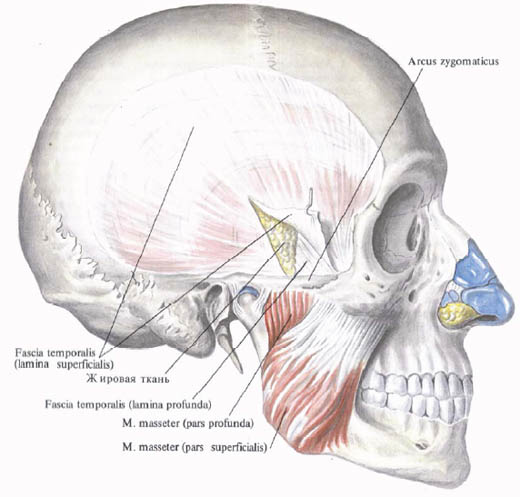
1. Chewing muscle , m. Masseter, originates from the lower edge of the zygomatic arch in two parts - superficial and deep.
The superficial part, pars superficialis, begins with tendon tufts from the anterior and middle sections of the zygomatic arch; The deep part, pars profunda, - from the middle and back sections of the zygomatic arch. Bunches of muscle fibers of the surface part follow obliquely down and back, deep - down and anteriorly. Both parts of the masticatory muscle are joined and attached to the outer surface of the mandibular branch and to its corner in the area of masticatory tuberosity.
Function: lifts the lowered lower jaw; The superficial part of the muscle participates in the extension of the jaw forward.
Innervation: n. Massetericus (n. Trigeminus).
Blood supply: aa. Masseterica, transversa faciei.

2. The temporal muscle , m. Temporalis, fills the temporal fossa, fossa temporalis. It starts from the temporal surface of the frontal bone of the large wing of the sphenoid bone and the scaly part of the temporal bone. Bunches of muscle, going down, converge and form a powerful tendon that passes through the inside of the zygomatic arch and attaches to the coronoid process of the lower jaw.
Function: the reduction of all muscle beams lifts the lowered lower jaw; The posterior tufts extended forward the lower jaw are pulled back.
Innervation: nn. Temporales profundi (trigeminus).
Blood supply: aa. Temporales profunda et superficialis.

3. The lateral pterygoid muscle , m. Pterygoideus lateralis, begins with two parts, or heads, the upper and lower.
The upper muscle head originates from the lower surface and from the trailing crest of the large wing of the sphenoid bone, is attached to the medial surface of the joint capsule of the temporomandibular joint and to the articular disk. The lower head starts from the outer surface of the lateral plate of the pterygoid process of the sphenoid bone and, guided backwards, is attached to the pterygoid pit of the lower jaw. Between the upper and lower head of the muscle there is a gap that passes through the buccal nerve.
Function: shifts the lower jaw in the opposite direction; Bilateral muscle contraction pushes the lower jaw forward.
Innervation: n. Pterygoideus lateralis (n. Trigeminus).
Blood supply: a. Alveolaris inferior (a. Maxillaris), a. Facialis.

4. The medial pterygoid muscle, m. Pterygoideus medialis, begins from the pterygoid pterygoid fossa, is directed back and down, attached to the pterygiform tuberosity of the mandible branch.
Function: shifts the lower jaw in the opposite direction; With a two-sided contraction, pushes forward and lifts the lowered lower jaw.
Innervation: n. Pterygoideus medialis (n. Trigeminus).
Blood supply: aa. Alveolares superior (a. Maxillaris), a. Facialis.


Comments
When commenting on, remember that the content and tone of your message can hurt the feelings of real people, show respect and tolerance to your interlocutors even if you do not share their opinion, your behavior in the conditions of freedom of expression and anonymity provided by the Internet, changes Not only virtual, but also the real world. All comments are hidden from the index, spam is controlled.