


Sphenoid bone
The sphenoid bone , os sphenoidale, unpaired, forms the central part of the base of the skull .
The middle part of the sphenoid bone is a body, corpus, of a cubic shape, has six surfaces. On the upper surface facing the cavity of the skull, there is a depression - the Turkish saddle, sella turcica, in the center of which is the pituitary fossa, fossa hypophysialis. It is pituitary, hypophysis. The size of the fovea depends on the size of the pituitary gland. The front of the Turkish saddle is the saddle, the tuberculum sellae. Behind him, on the lateral surface of the saddle, is the inconstant middle inclined process, processus clinoideus medius.
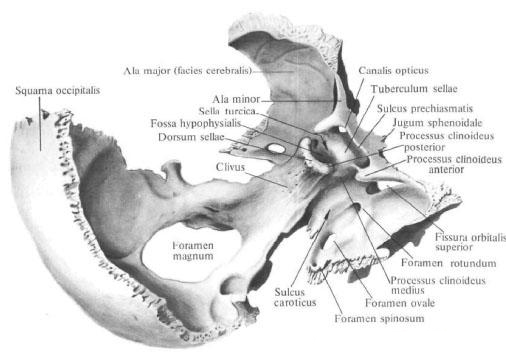
A shallow transverse pre-cross groove, sulcus prechiasmatis, extends from the hillock of the saddle. Behind her lies the cross of the optic nerves , chiasma opticum. The lateral furrow passes into the visual canal, canalis opticus. In front of the furrow is a smooth surface - a wedge-shaped elevation, jugum sphenoidale, connecting the small wings of the sphenoid bone. The front faucet of the upper surface of the body is jagged, slightly protruding and joins with the posterior edge of the trellis of the trellis, forming a wedge-latticed suture, sutura spheno-ethmoidalis. The back border of the Turkish saddle is the back of the saddle, dorsum sellae, which ends right and left with a small posterior inclined processus, processus clinoideus posterior.
On each side of the saddle, a carotid sulcus (a trail of the internal carotid artery and the accompanying plexus) follows from behind. At the posterior edge of the furrow, from its outer side, is a pointed process - a wedge-shaped tongue, lingula sphenoidalis.
The posterior surface of the saddle back passes into the upper surface of the basilar part of the occipital bone , forming a ramp, clivus (on it lie the bridge, the medulla oblongata, basilar artery and its branches). The posterior surface of the body is rough; Through the cartilaginous layer it joins the anterior surface of the basilar part of the occipital bone and forms a wedge-occipital synchondrosis, synchondrosis spheno-occipitalis. With age, cartilage is replaced with bone tissue and both bones coalesce.
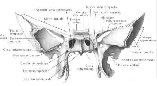
The anterior surface of the body and part of the lower surface are facing the nasal cavity. In the middle of the front surface there is a wedge-shaped crest, crista sphenoidalis; Its anterior margin is attached to the perpendicular plate of the trellis. The lower process of the ridge is pointed, extends downwards and forms a wedge-shaped beak, rostrum sphenoidale. The latter connects with the vomer's wings, alae vomeris, forming a vomer-beak-like canal, canalis vomerorostratis, lying along the middle line between the upper edge of the opener and the wedge-shaped beak. Lateral from the ridge lie thin curved plates - wedge-shaped conchae, conchae sphenoidales. The shells form the anterior and partly the inferior walls of the sphenoid sinus, sinus sphenoidalis. In each shell there is a small aperture - aperture of the sphenoid sinus, apertura sinus sphenoidalis. Outside the aperture, there are insignificant cavities that cover the cells of the posterior part of the maze of the latticed bone. The outer edges of these indentations partially connect with the orbital plate of the trellis, forming a wedge-latticed suture, sutura spheno-ethmoidalis, and the lower ones with the orbital processes, orbitalis, palatine bone.

The sphenoid sinus, sinus sphenoidalis, is a pair of cavities that occupy most of the body of the sphenoid bone; It refers to airborne paranasal sinuses. The right and left sinuses separate one from another the septum sinuum sphenoidalium. Which extends anteriorly into a wedge-shaped crest. As in the frontal sinuses, the septum is often asymmetric, as a result of which the size of the sinuses may not be the same. Through the aperture of the sphenoid sinus, each sphenoid sinus communicates with the nasal cavity. The cavity of the sphenoid sinus is lined with a mucous membrane.
Small wings, alae minores, the sphenoid bone extend in both directions from the anterolateral corners of the body in the form of two horizontal plates, at the base of which there is a rounded hole. From this hole begins the bone canal up to 5-6 mm in length - the visual canal, canalis opticus. The optic nerve lies in it, n. Opticus, and the ocular artery, a. Ophthalmica, Small wings have an upper surface facing the cranial cavity, and a lower one that points into the orbit cavity and closes the upper glandular fissura, fissura orbitalis superior.
The anterior edge of the small wing, thickened and serrate, joins the orbital part of the frontal bone . The posterior margin, concave and smooth, freely protrudes into the cavity of the skull and is the boundary between the anterior and middle cranial fossae, fossae cranii anterior et media. Medially the posterior edge ends with a protruding, well pronounced anterior inclined processus, clinoideus anterior (a portion of the dura mater is attached to it - the diaphragm of the Turkish saddle, diaphragma sellae).
Large wings, alae majores, extend from the lateral surface of the body of the sphenoid bone and are directed to the outside.
The large wing has five surfaces and three edges. The upper cerebral surface, facies cerebralis, concave, faces the cavity of the skull. It forms the anterior section of the middle cranial fossa. It is distinguished by finger impressions, impressiones digitatae [gyrorum], and arterial furrows, sulci arteriosi (impressions of the relief of the adjacent surface of the brain and middle meningeal arteries).
At the base of the wing there are three permanent holes: inside and anteriorly there is a round hole, foramen rotundum (through it comes the maxillary nerve, n maxillaris); Outside and behind the round there is an oval hole, foramen ovale (it passes the mandibular nerve, n. Mandibularis), and outside and behind the oval - a spinous hole, foramen spinosum (through it come the middle meningeal artery, vein and nerve). In addition, in this region there are non-permanent holes. One of them is a venous aperture, foramen venosum, located somewhat posteriorly from the oval aperture. It misses a vein, going from the cavernous sinus to the pterygoid venous plexus. The second is a stony hole, foramen petrosum, through which passes a small rocky nerve, a winged suture, sutura sphenofrontalis. The outer divisions of the frontal margin terminate with a sharp parietal margin, margo parietalis, which, with a wedge-shaped angle of the other bone, forms a wedge-parietal suture, sutura sphenoparietalis. The inner parts of the frontal margin go into a thin free edge that is separated from the lower surface of the small wing, bounding the upper globule from below.
Anterior zygomatic margin, margo zygomaticus, serrate. The frontal process, processus frontalis, zygomatic bone and zygomatic margin join together to form a wedge-zygomatic suture, sutura sphenozygomatica.
The posterior scaly margin, margo squamosus, connects with the wedge-shaped margin, margo sphenoidalis, the temporal bone and forms a wedge-scaly seam, sutura sphenosquamosa. Behind and outward, the scaly edge ends with the spine of the sphenoid bone (the place of attachment of the wedge-mandibular ligament, lig sphenomandibularis, and muscle bundles straining the palatal curtain, M. tensor veli palatini).
Inside the spine of the sphenoid bone, the posterior edge of the large wing lies in front of the stony part, the pars petrosa, the temporal bone and bounds the wedge-stony fissure, fissura sphenopetrosa, medially turning into a torn hole, foramen la-lacerum; On an uncertified skull this gap is made of a cartilaginous tissue and forms a wedge-stony synchondrosis, synchondrosis sphenopetrosa.
The pterygoid processes, processus pterygoidei, move away from the junction of the large wings to the body of the sphenoid bone and are directed downward. They are formed by two plates - lateral and medial. Lateral plate, lamina lateralis (processus pterygoidei), wider, thinner and shorter than the medial (from its outer surface begins the lateral pterygoid muscle, m. Pterygoideus lateralis).
The medial plate, lamina medialis (processus pterygoidei), is already thicker and slightly longer than the lateral. Both plates fuse with their anterior edges and, spreading posteriorly, border the pterygoid fossa, fossa pterygoidea (here begins the medial pterygoid muscle, m. Pterygoideus medialis). In the lower trimmed
Both plates do not fuse and border the pterygium, incisura pterygoidea. In it is located pyramidal process, processus pyramidalis, palatine bone. The free end of the medial plate ends in a downward and outward-directed pterygoid hook, hamulus pterygoideus, on the outer surface of which there is a furrow of the pterygoid hook, sulcus hamuli pterygoidei (through which the tendon of the muscle straining the palatal curtain, M. tensor veli palatini) is thrown.
The posterior margin of the medial plate at the base widens and forms a scaboid fossa about the share of the cotton-shaped fossa, the fossa scaphoidea.
Outside the scaphoid fossa is a shallow furrow of the auditory tube, sulcus tubae auditivae, which laterally passes to the lower surface of the posterior edge of the large wing and extends to the spine of the sphenoid bone (a cartilaginous part of the auditory tube extends to this furrow). Above the navicular fossa and medial there is an opening with which the canopy canal begins, canalis pterygoideus (vessels and nerves pass through it).
The canal extends sagittally in the thickness of the base of the pterygoid process and opens on the maxillary surface of the large wing, on the posterior wall of the pterygopalatine fossa.
The medial plate at its base passes into a flat, horizontally moving vaginal process, a processus vaginalis, which is located under the body of the sphenoid bone, covering the vomer wing, ala vomeris. At the same time, the groove of the vaginal process facing the wing of the vomer is the vomerine-vaginal furrow, sulcus vomerovaginalis, turns into the vomer-vaginal canal, canalis vomerovaginalis.
Outside the appendage, there is a sagittally running small, crescent-vaginal groove, sulcus palatovaginalis. The contiguous wedge-shaped process of the palatine bone, processus sphenoidalis ossis palatini, closes the furrow in the eponymous canal, canalis palatovaginalis (in the vomerine-vaginal and neural-vaginal canals pass the nerve branches of the pterygoid node, and in the vaginal-vaginal canal, in addition, the wedge-palatal Arteries).
Sometimes, from the posterior edge of the outer plate toward the spine of the sphenoid bone, the pterygoid-spinous process, the processus pterygospinosus, which can reach the indicated awn and form an aperture, is directed.
The anterior surface of the pterygoid process connects with the posterior surface of the upper jaw in the region of the medial edge of the tuber, forming a wedge-maxillary suture, sutura sphenomaxillaris, which lies in the depth of the pterygopalatine fossa.
You will be interested to read this:

Comments
When commenting on, remember that the content and tone of your message can hurt the feelings of real people, show respect and tolerance to your interlocutors even if you do not share their opinion, your behavior in the conditions of freedom of expression and anonymity provided by the Internet, changes Not only virtual, but also the real world. All comments are hidden from the index, spam is controlled.