


Kidneys
Kidney , gep, - paired bean-shaped organ. The kidneys are located in the abdominal cavity, in the lumbar region, on either side of the spine. Each kidney reaches a length of 10-12 cm, a width of 5-6 cm, its thickness is about 4 cm. The mass of one kidney is 120-200 g. The left kidney is somewhat longer than the right kidney, sometimes it has a large mass. The color of the kidneys is often dark brown.
In the kidney, the front and back surfaces, the lateral and medial margins, the upper and lower ends are distinguished.

Anterior surface, fades anterior, convex and slightly laterally oriented. Upper% of the right kidney is adjacent to the liver, and the upper third of the left kidney is to the stomach. The posterior surface, fades posterior, is flattened. The side portion of each kidney is attached to the square muscle of the waist. The lateral margin, margo lateralis, is convex and is directed somewhat towards the back wall of the stomach; The medial margin, margo medialis, is concave and faces downward, medially and forward.
In the middle of the medial edge of the kidney there is a depression - the renal gates, hilum renalis, which passes into the renal sinus, sinus renalis. The portal of the kidney is bounded by two projections of the medial margin, of which the posterior protrusion protrudes. As a result, the posterior surface of the kidney is wider than the anterior, and the renal sinus is more anterior to the anterior.
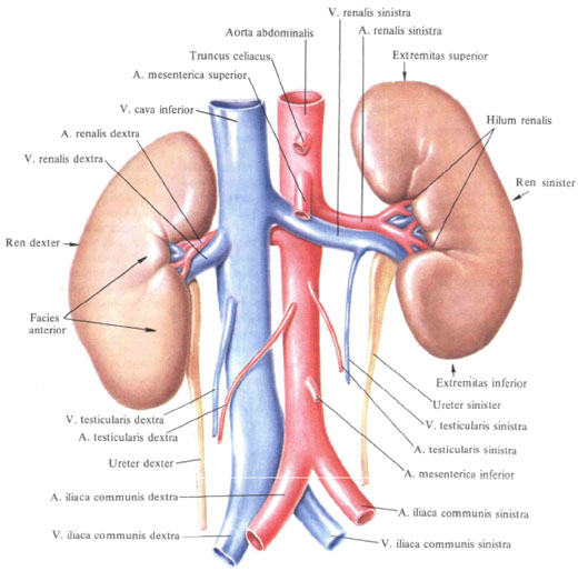
In the renal sinus are the renal pelvis, pelvis renalis, renal cups, calices renales, renal vascular and nerve branches, lymph nodes and fatty tissue. The mutual arrangement of the listed entities entering the gates is such that the veins lie in front, arteries and nerves behind the veins, and the renal pelvis and ureter are posterior to the arteries.
The upper end, extremitas superior, the kidneys are wider than the lower extremitas inferior. At the upper ends of the adrenal glands, glandulae suprarenales. These ends are closer to the median plane of the body than the lower ones; The latter are more deviated from the spinal column.
The kidneys are covered with a dense fibrous capsule, capsula fibrosa, which consists of the outer connective tissue layer and the internal smooth muscle; Fibers of smooth muscles penetrate into the tissue of the kidney. The capsule is weakly fused to the substance of a healthy kidney, and if you make an incision on it, it is easy to remove.
Each kidney is surrounded by a fatty capsule, and outside - a renal fascia. The fat capsule, capsula adiposa, directly envelops the kidney, covering the thicker layer with its posterior surface; Through the kidneys it penetrates into the renal sinus.

Renal fascia, fascia renalis, represents part of the retroperitoneal fascia,
At the lateral edge of the kidney is divided into two plates: the anterior, or the preference, and the posterior or backbone, the plates that surround the kidney along with the fatty capsule, as well as the adrenal gland, kidney vessels and nerves located at the upper end of the kidney. Medial to the kidney, the posterior fascia leaf extends over the surface of the vertebral bodies; The front leaf passes in front of the large vessels of the abdominal cavity: the inferior vena cava and the abdominal aorta into the anterior plate of the renal fascia of the opposite side. Towards the upper end of the kidney, both plates of the renal fascia merge; Down they do not unite and pass into the sub-peritoneal tissue of the iliac fossa. The fat capsule is pierced by connective tissue strands that extend from the renal fascia to the fibrous capsule of the kidney.
On the anterior surface of the renal fascia is the perineum fat body, corpus adiposum pararenals.
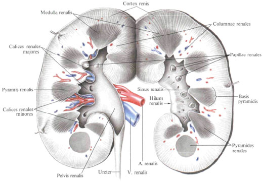
On the cut of the kidney it can be seen that it consists of a cerebral and cortical substance that differ in density and color: the brain substance is denser, bluish-red, cortical - yellowish-red; These differences depend on the unequal blood supply. The cerebral substance occupies the central part of the organ, the cortical is its periphery.
The brain substance, medulla renalis, is not a continuous mass, but consists of cone-shaped formations - kidney pyramids, pyramides renales, whose number reaches 15-20 or more. The base of each pyramid, the basis of pyramidis, faces the outer surface of the kidney, the apex is directed toward the sinus.
Cortex, cortex renais, has a thickness of 5-7 mm, it seems to border the convex base of the pyramids and gives between them processes directed towards the center of the kidney - kidney posts, columnae renales. The cortical substance consists of two parts: the radiant part, the pars ra-diata, and the folded part, the pars convoluta. The radiant part is a continuation of the medullary substance from the base of each renal pyramid. The curled part is a segment of the cortical lobule, consisting of renal corpuscles and proximal and d and steel tubules of the nephron, lying between the rays. In the embryonic period and in early childhood, the pyramids with the surrounding cortex, the so-called papal lobes, tobi rcnales, are clearly visible. In these periods, the kidney looks lobed. With age, the borders between the lobes gradually become smoother, while in the cortical substance there are signs of lobulation in the form of cortical lobuti corticales.
The tops of the pyramids, merging 2-3 (sometimes up to 6), form renal papillae protruding into the renal sinus, papillae renales. At the top of the papilla are papillae, foramina papillaria, from 10 to 55, forming a latticed field, area cribrosa, papilla. The renal papillae are covered with funnel-shaped small kidney cups, calices renales minores, whose number on average reaches 8-9; Sometimes one small cup covers 2 or even 3 papillae. Several small kidney cups are connected to a large kidney cup, ealix renal is major; Their 2-4, they are essentially urinary ducts that connect individual groups of small kidney cups to the renal pelvis.

The renal pelvis, pelvis renalis, has a narrowed shape in the anteroposterior direction of the funnel; Its broad part is embedded in the sinus, while the narrowed part protrudes outward in the area of the kidneys' gate and passes into the ureter. The cavities of small and large cups are lined with mucous membrane, which directly passes into the mucous membrane of the pelvis, and the latter - into the mucous membrane of the ureter.
Functionally the most important part of the renal tissue is the epithelial ducts - urinary renal tubules, lubuti renales. They take part in the formation of the structural and functional unit of the kidney - not a ponen, nenhro-pit, consisting of a renal corpuscle and a system of nephron tubules. The kidney-
Uf \ O? Γ \ ΡΛ.. \ С
From the vascular glomerulus - the glomerulus of the renal corpuscle, glomerulus corpusculi re-nalis, and the two-layered glomerulus capsule, capsula glomeruli,

The renal (urinary) tubule escaping from the capsule is in the cortical substance of the kidney the proximal part of the nephron tubule, pars proximo lis tubuli nephron !, passing into the nephron nepron, ansa nephron !. The nephron loop lies in the brain substance of the kidney. In it, a descending
Part, pars descendens ansae, and the ascending part, pars ascendens ansae, passing into the distal spicy canaliculus
Uptfinciutt n / irv / tietnlie tithnli illD * rj Qg. '* * "J * y **»' ^ i ^ »'LJ UIMIWIM IKUUII in until until, rt Jfl»
Those into the collective renal tubules, tubuli renales colligenies. A few cotypsis trips are inserted into the papillary ducts, ductus papillaris, the latter are buried with co-osaceous holes, foramina paptl-laria, on the lattice field, area cribrosa, renal papilla, papilla renalis, on top of the pyramid renum, pyramis renalis.
Blood vessels are particularly closely related to the renal tubule system. Branches of the renal artery, a. Renalis. Penetrating from the renal sinus, sinus renalis, into the renal substance, spread out radially between the pyramids in the form of the interlobar arteries of the kidney, aa. In-terlobares rents.
APPLYING TO CAPITAL? CORK AND
Of the medulla, each interlobar artery is divided into two arterial arteries, aa. Arcuatae. Entering into neighboring parts and located here above the base of the pyramid. They send spiral arterioles to the brain substance. Arleriolae rectae, and into the cortical substance - interlobular arteries, aa. Inieriobulares, terminating in a fibrous capsule with capsular branches. N. Capsulars.
From the interlobular arteries, the glomerular arterioles (bringing the blood vessels), arteriole glomerularis afferens (vas afferens) depart. Which decay-
On the capillary nodule, rete capillare glomerular, surrounded by a capsule. The capillary network is only arterial (by the type of a wonderful network, rete mtrabile), and the glomerular blood capillary, vas haemocapillare glomerular, emerging from the glomerular network, passes into the outgoing club-arteriol (vasoconstrictor), arteriola glomerularis efferens (uas efferens) Outside the capsule. This capillary again breaks up into a network of capillaries, braiding the urinary tubules and giving rise to the venous system.
From the brain substance, blood is collected from the spicy venules, venulae rcciae, flowing into the arched veins, vv. Arcuatae. In the cortex, according to the course of the interlobular arteries, there are memsolporic veins. about". Inlerfabulares. The latter are formed from small vessels of the surface layer of the cortico-
Tm * 1rn R ^ mprTRa t'kl iraTumnp \ f ^ rv iapif \ un-
Th veins, vv. Stellatae. And in the future take the veins from the secondary capillary network, braiding the renal tubules. Interlobular veins are poured into the arched veins. The arc veins of two adjacent lobes, merging, form the interlobar veins, vv. Intertobares, which follow through the renal columns along with the interlobar arteries. In the circumference of the renal papillae, the interlobar veins emerge from the kidney parenchyma into the renal sinus, where, merging, form a renal vein, v. Renalis, which empties into the inferior vena cava, v. Cava inferior. In each kidney, according to the division of the artery, the renal segments, tegmenta renalia (see Figure 628), are distinguished: the upper segment, segmentum
SlinerklS, COOTBeTCTBve-g medial
The edge and partly the anterior surface of the upper end of the kidney; The upper front segment, segmentum anlcrhts supc rius, includes the front surface
Upper end, upper part of middle part of kidney, lateral margin and partly posterior surface; Lower anterior segment, segmentum anterius
MforistQ IPWUT vnf npnYiriiH npnpnmiff
Segment, in front of the renal pelvis, reaching the anterior surface of the kidney in the lower part of its middle part and partly on the posterior surface; The lower segment, segmentum infe-rius, occupies the lower end of the night; Posterior segment, segmentum posterius, lies behind the renal pelvis and corresponds to the posterior surface of the kidney between the upper segment from the top. Bottom - bottom, upper and lower anterior segments - laterally. Shintoia and skeleotopia of the right and left kidneys are different. The right kidney is located from the XII thoracic to the upper edge of the IV lumbar vertebra, the left kidney from the XI thoracic to the upper edge of the 3rd lumbar vertebra. The female kidneys are wi. V-vertebrae lower than that of men. The width of the kidneys is from the lateral edge of the large lumbar muscle to the posterior margin of the transverse abdominal muscle. The back surface
Both buds are adjacent to the diaphragm (upper stumps): the remaining areas of the surface are adjacent: medially to the lumbar muscle, laterally - to the square. Gshyashshshy and transverse abdominal muscle. Both buds are located in front of the XII rib, which runs relatively long along their axis obliquely from the top and from the outside; The right kidney is intersected by the XII edge at the border of its upper and middle third, its uppermost part only reaches the XI edge: the left kidney is intersected by the XII rib almost in the middle of the length, and the upper outer segment is located somewhat higher than the level of the XI edge.
Directly to the upper shock of the right kidney is adhered to the right adrenal gland (see Figure 615). The right side of the liver contacts the anterior surface of the right kidney during the upper ~ A; Lower liver to the anterior surface of the right kidney is the right bend of the colon: adjoining the meadalymium \ portion and the ores is the descending part of the duodenum. The anterior surface of the right kidney is covered with the peritoneal gland only at the site of contact with the liver.
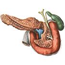
The upper end of the left kidney is adhered to the left adrenal gland; In the area of the upper third, the anterior surface of the left kidney touches the back wall of the stomach, in the middle third - with the tail of the pancreas, which crosses the gate of the kidney in the transverse direction. To the lateral margin of the left kidney, over its upper half, the spleen is due. The lower third of the antero-
Mрлматтирпгп vuarTi / a ГТРЕ / ЛН nnuiMi лЛ-
AND I | -.- L G L LI. IUIIU1 Vy T tUV L No.U OUUVK AI \ J 1 IS 4 VVV
Is wound in the left mesenteric sinus and comes into contact here with the loins of the jejunum, the left flexure of the colon is located along the anterior part of the left kidney. The anterior surface of the left kidney in the areas of contact with the stomach, spleen and jejunum is covered with the peritoneum.
The kidneys are susceptible to various anomalies of development and position. Particularly variable is the position of the right kidney, due to the lowering of the colon. Sometimes, instead of two, there is one kidney located in the pelvis; In some cases, there is an arched or horseshoe-shaped kidney - the kidneys fused with the lower ends.
Innervation: plexus renalis (derivative of plexus celiacus), is formed by the branches of vagus and truncus sympathicus. Sensitive innervation is carried out from the lower thoracic and upper lumbar spinal nodes.
Blood supply: a. Renalis (aorta abdominalis branch). Venous blood flows out by v. Renalis in v. Cava inferior. Lymphatic vessels carry lymph to the nodi lymphatici lumbales.


Comments
When commenting on, remember that the content and tone of your message can hurt the feelings of real people, show respect and tolerance to your interlocutors even if you do not share their opinion, your behavior in the conditions of freedom of expression and anonymity provided by the Internet, changes Not only virtual, but also the real world. All comments are hidden from the index, spam is controlled.