


Angiology - the doctrine of the vessels
Circles of blood circulation
A heart
- External structure of the heart
- The cavity of the heart
- Right atrium
- Right ventricle
- The left atrium
- Left ventricle
- Heart wall structure
- Conductive system of the heart
- Heart vessels
- Topography of the heart
- Pericardium
Small vessels of the circulatory system
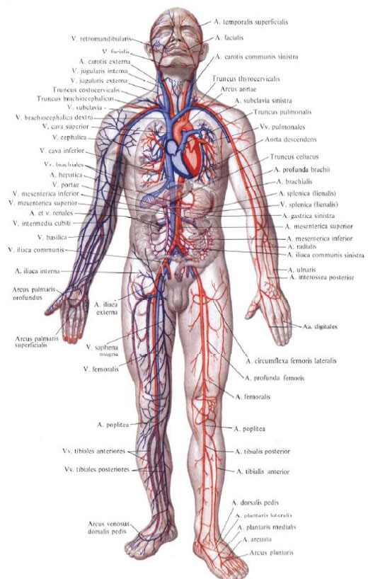
Arteries of a great circle of blood circulation
Arteries of the upper limb
Torso arteries
- The thoracic part of the aorta
- Abdominal part of the aorta
- Common iliac artery
- Internal iliac artery
- External iliac artery
Arteries of the lower limb
Given a number of morphological and functional characteristics, a single vascular system is divided into the circulatory system, systema Sanguineum , and lymphatic system, systema Limphaticum . The vascular system that transports blood, haema, and lymph, is closely related to the system of hematopoietic and immune organs (bone marrow, thymus, lymph nodes, lymphoid tissue of palatine, lingual, tubal and other tonsils, spleen and liver - in the embryonic period) Constantly replenishing the perishing elements of blood.
In accordance with the direction of movement of blood, blood vessels are divided into arteries, arteriae , which bring blood from the heart to the organs, capillaries, vasa sar illaria , through the wall of which metabolic processes occur, and veins, venae , - vessels carrying blood from organs and tissues to the heart .
The arteries are successively branched into smaller vessels with thinner walls. Their smallest branches are arterioles, arteriolae , and precapillaries, precapillares , passing into capillaries. Of the latter, blood collects into postcapillaries, postcapillares , and further into venules, venulae , connecting into small veins. Arterioles, precapillaries, capillaries, postcapillaries, venules, as well as arteriolovenous anastomoses, anastomoses arteriolovenulares , constitute a microcirculatory bed that provides a metabolism between blood and tissues in the organs. The microcirculatory bed also includes lymphocapillary vessels, vasa Lymphocapillares , the spatial position of which is closely related to the blood capillaries.
The structure of the microcirculatory bed depends on the type of branching of the arterioles.
For arcade type branching of arterioles is characterized by the formation of numerous anastomoses between their branches, as well as between the tributaries of the venules. At terminal type of branching of arterioles anastomoses between terminal branches of arterioles are not formed: after branching by several orders, arterioles without a sharp border pass into precapillaries, and the latter into capillaries. The structure of the microcirculatory bed is characterized by pronounced organ-specific features, which are caused by the specialization of the blood capillaries.
Walls of arteries, veins and lymphatic vessels consist of three layers: internal, middle and outer.
Inner shell, tunica Intima , the vessel consists of the endothelium represented by closely adjacent endotheliocytes located on the subendothelial layer, which is cambial for the latter.
Middle shell, tunica Media , is formed mainly by circularly located smooth muscle cells, as well as connective tissue and elastic elements.
Outer shell, tunica Externa , consists of collagen fibers and a series of longitudinal bundles of elastic fibers.
Blood vessels, both blood and lymphatic, are supplied with blood, small thin arteries and veins - vessels of vessels, vasa Vasorum , and lymph flows off the lymph vessels of the vessels, vasa Lymphatica Vasorum .
Vascular neural plexuses located in the outer and middle shells of the vessel wall and formed by the nerves of the vessels provide the innervation of the vessels. Vasorum . The composition of these nerves includes both vegetative and somatic (sensitive) nerve fibers.
The structure of the walls of arteries and veins is different. The walls of the veins are thinner than the walls of the arteries; The muscle layer of veins is poorly developed. In the veins , especially in the small and medium, there are venous valves, valvulae Venosae .
Depending on the degree of development of the muscular or elastic elements of the middle shell, the arteries of the elastic type (aorta, pulmonary trunk), muscular-elastic type (carotid, femoral and other arteries of the same caliber) and arteries of the muscular type (all other arteries) are distinguished.
The walls of the capillaries consist of a single layer of endothelial cells located on the banal membrane.
The caliber and thickness of the walls of blood vessels as they are removed from the heart as a result of gradual division in the organs and tissues of the body change. In each organ the character of branching of vessels, their architectonics, have their own peculiarities.
Outside and intraorganic vessels, joining together, form anastomoses, or anastomoses (extraorganic and intragroup). In some places the anastomoses between the vessels are so numerous that they form the arterial network, rete Arteriosum , Venous network, rete Venosum , or vascular plexus, plexus Vasculosus . By means of anastomoses, more or less removed from each other parts of the vascular trunk, as well as vessels in the organs and tissues. These vessels take part in the formation of collateral (roundabout) blood circulation (collateral vessels, vasa Collateralia ) and can restore blood circulation in this or that part of a body at difficulty of movement of blood along the main trunk.
In addition to anastomoses connecting two arterial or venous vessels, there are connections between arterioles and venules - these are arteriolovenous Anastomoses, anastomoses Arteriolovenulares . Arteriolovenous anastomoses form the so-called apparatus of reduced blood circulation - the derivational device.
In some parts of the arterial and venous system there is a wonderful network, rete Mira b ile . It is a network of capillaries in which the delivering and vasodilating vessels are the same: for example, in the glomerulus of the renal corpuscle, glomerulus renalis, where the bringing arterial vessel is divided into capillaries that reconnect to the arterial vessel.
Angiology, angiologia
(From the Greek angeion - vessel and logos - teaching), combines data on the study of the heart and the vascular system.

Comments
When commenting on, remember that the content and tone of your message can hurt the feelings of real people, show respect and tolerance to your interlocutors even if you do not share their opinion, your behavior in the conditions of freedom of expression and anonymity provided by the Internet, changes Not only virtual, but also the real world. All comments are hidden from the index, spam is controlled.