


Vesicular duct
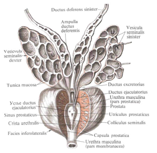
The vas deferens, ductus deferens, paired, is a dense tube up to 50 cm long, 3.0-3.5 mm in diameter with a lumen of 0.5-0.7 mm. The duct starts from the lower end of the appendage tail and opens with a common duct with a seminal vesicle into the prostate part of the urethra.
Topographically, the seminiferous duct consists of several sections. The initial section, located along the posterior edge of the testicle, represents a sharply convoluted cylindrical tube (the testicle part of the duct). The second part of it goes in the spermatic cord (the cord part) in the scrotum, reaches the superficial inguinal ring and lies further in the inguinal canal (inguinal part). Going out through the deep inguinal ring, the vas deferens along the side wall of the small pelvis (the pelvic part), resting in the retroperitoneum, and reaches the bottom of the bladder. In these areas, it looks like an even cylindrical tube of white color. In the pelvic part, the duct has an extension - the ampulla of the vas deferens, ampulla ductus deferentis, its wall here represents bay-like cavities - ampulla diverticula, diverticulae ampullae, visible from the outside in the form of tuberosity.

The terminal part of the vas deferens again tapers. By connecting with the excretory duct, ductus excretorius, the seminal vesicle, it forms the ejaculatory duct, ductus ejaculatorius.
In the wall of the vas deferens ducts are distinguished: the outer, the adventitial, the middle - the muscular and the inner - the mucous membrane. The adventitia, tunica adventitia, consists of connective tissue fibers and a small amount of elastic fibers. In it pass feeding vessels and nerve elements of the duct. The muscular membrane, tunica muscularis, is the thickest part of the wall and consists of the outer and inner longitudinal layers and the middle circular layer of smooth muscles. The mucous membrane, tunica mucosa, forms longitudinal folds; It is covered with a multi-row prismatic epithelium. In its own connective tissue plate there is a significant amount of elastic fibers.
Inversion: sympathetic and parasympathetic nerves, plexus deferentialis (from the plexus hypogastrics inferior).
Blood supply: ascending branch a. Ductus deferentis (from a. Umbilicalis), ampulla of the vas deferens - a. Rectalis media, a. Vesicalis inferior and descending branch a. Ductus deferentis. Venous blood flows down the accompanying duct vein flowing into the plexus venosus vesicalis and then to v. Iliaca interna. Lymphatic vessels carry lymph in the nodi lymphatici iliaci interni.
You will be interested to read this:

Comments
When commenting on, remember that the content and tone of your message can hurt the feelings of real people, show respect and tolerance to your interlocutors even if you do not share their opinion, your behavior in the conditions of freedom of expression and anonymity provided by the Internet, changes Not only virtual, but also the real world. All comments are hidden from the index, spam is controlled.