


Uterus
The uterus, uterus (metra), represents an unpaired hollow smooth muscle organ located in the pelvic cavity, at the same distance from the pubic symphysis and sacrum , at such an elevation that the uppermost portion of the uterus does not protrude beyond the level of the upper aperture of the pelvis. Uterus pyriform, flattened in anteroposterior direction. The wide part of it is facing upwards and anteriorly, narrow - downwards. The shape and size of the uterus varies significantly during different periods of life and mainly in connection with pregnancy. The length of the uterus in a nulliparous woman is 7-8 cm, in those giving birth, 8-9.5 cm, the width at the bottom is 4-5.5 cm; Mass varies from 30 to 100 g.
In the uterus, the neck, body and bottom are distinguished.
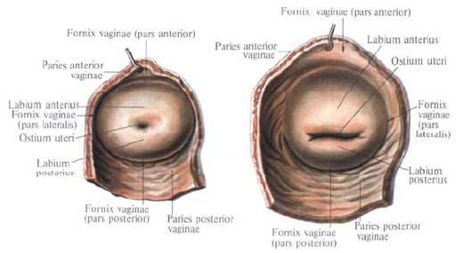
The cervix of the uterus , cervix uteri, sometimes gradually passes into the body, sometimes sharply separating from it; Its length reaches 3-4 cm; It is divided into two parts: supravaginal and vaginal. The upper two thirds of the cervix is located above the vagina and make up its supra-vaginal part (cervix), portio supravaginalis (cervicis). The lower part of the cervix is pressed into the vagina and constitutes its vaginal part, portio vaginalis (cervicis). At its lower end there is a round or oval aperture of the uterus, ostium uteri, whose edges form the anterior lip, labium anterius, and the posterior lip, labium posterius. In women giving birth, the hole of the uterus has the form of a transverse slit, in nulliparous - round. The posterior lip is somewhat longer and less thick, located above the anterior. The opening of the uterus is directed towards the back wall of the vagina.
In the region of the cervix, there is a cervical canal, canalis cervicalis uteri, whose width is not the same throughout: the middle sections of the channel are wider than the area of the outer and inner openings, as a result of which the channel cavity is fusiform.
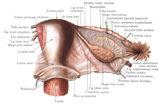
The body of the uterus, corpus uteri, has the shape of a triangle with a truncated lower corner extending into the cervix. The body is separated from the neck by the narrowed part - the isthmus of the uterus, isthmus uteri, which corresponds to the position of the inner orifice of the uterus. In the body of the uterus, the anterior vesicle surface is distinguished, the facies vesicalis, the posterior intestinal surface, the facies intestinalis, and the lateral, right and left margins of the uterus, the margins uteri (dexter et sinister), where the anterior and posterior surfaces pass into each other. The upper part of the uterus, which rises in the form of a vault above the orifices of the fallopian tubes, is the bottom of the uterus, the fundus uteri. With the lateral edges of the uterus, the uterus's bottom forms corners, into which the fallopian tubes enter. The section of the body of the uterus, corresponding to the place of the confluence of the tubes, is called the uterine horns, cornua uteri.
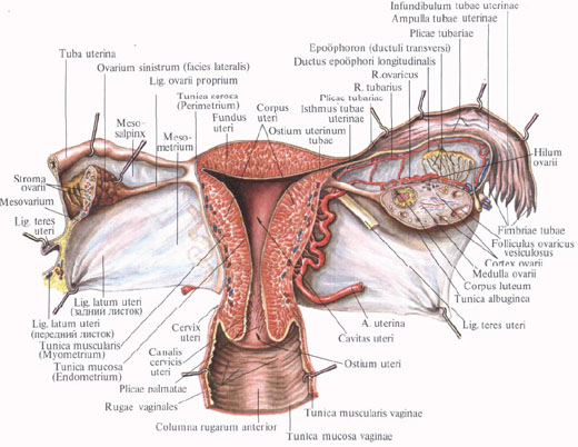
The cavity of the uterus, cavitas uteri, 6-7 cm in length, is triangular in the frontal incision, in the upper corners of which the oviducts are opening, in the lower corner there is an internal hole of the uterus that leads to the cervical canal. The size of the cavity of the nulliparous is different than that of those giving birth: the first side walls are more sharply concave into the cavity. The anterior wall of the uterus body adjoins the posterior wall, whereby the cavity on the sagittal incision is in the form of a slit. The lower narrow part of the cavity communicates with the cervical canal, canalis cervicis uteri.
The wall of the uterus consists of three layers: the outer layer - the serosa, the tunica serosa (perimetrium), the subserous base, tela subserosa, the middle - muscular, tunica muscularis (myometrium), and the inner - mucosa, tunica mucosa (endometrium).
Serous membrane (perimetry), tunica serosa (perimetrium), is a direct continuation of the serous cover of the bladder. On a large extent of the anterior and posterior surfaces and the bottom of the uterus, it is through a subserous base, tela subserosa, tightly fused to the myometrium; At the border of the isthmus, the peritoneum cover is loose.
The muscular membrane of the uterus (myometrium), tunica muscularis (myometrium), the most powerful layer of the uterine wall, consists of three layers of smooth muscle fibers with an admixture of loose fibrous connective tissue. All three layers are intertwined with their muscle fibers in various directions, and as a result, the separation into layers is not well expressed. A thin outer layer (subserosal) consisting of longitudinally arranged fibers and a small number of circular (circular) fibers, is closely fused with the serous cover. The middle layer, circular, is most developed. It consists of muscle beams forming rings that are located in the region of the tube angles perpendicular to their axis, in the region of the uterus body - in a circular and oblique directions. This layer contains a large number of vessels, mainly venous, so it is also called the vascular layer, stratum vasculosum. The inner layer (submucosa) is the thinnest, with longitudinally extending fibers.
The mucous membrane of the uterus (endometrium), tunica mucosa (endometrium), fusing with the muscular membrane, lining the uterine cavity without a submucosa base and passes to the openings of the fallopian tubes; In the region of the bottom and body of the uterus, it has a smooth surface. On the anterior and posterior walls of the cervical canal, the mucosa, endocervix, forms longitudinally reaching palmoid folds, plicae palmatae. The mucous membrane of the uterus is covered with a single-layer prismatic epithelium; It contains simple tubular uterine glands, glandulae uterinae, which in the neck region are called cervical glands (cervical uteri), glandulae cervicales (uteri).
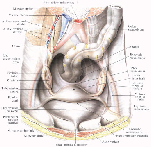
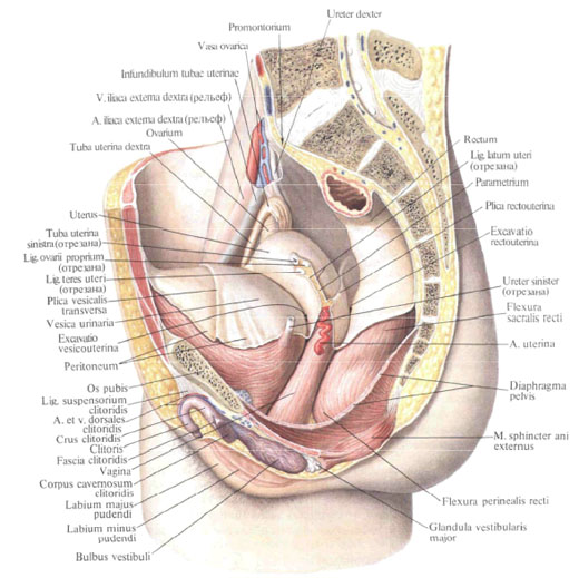
The uterus occupies a central position in the pelvic cavity. Anterior to it, in contact with its front surface, is the bladder, behind - the rectum and the loops of the small intestine. The peritoneum covers the anterior and posterior surfaces of the uterus and passes to the neighboring organs: the bladder, the front wall of the rectum. On the sides at the site of the transition to the broad ligaments, the peritoneum is connected with the uterus loosely. At the base of the broad ligaments, at the level of the cervix, between the leaves of the peritoneum is located the ovary cellulose, or parametrium, passing into the cervical region of the cervix paracervix.
The lower half of the front surface of the cervix is devoid of serous cover and is separated from the upper part of the posterior wall of the bladder by a connective tissue membrane, fixing both organs to each other. The lower part of the uterus - the cervix - is fixed to the vagina that starts from it.
The uterus occupies in the cavity of the small pelvis not a vertical, but an anteriorly curved position, anteversio, as a result of which its body is tilted over the anterior surface of the bladder. The body of the uterus along the axis forms an angle 70-100 ° open to the anterior to the anterior neck - an anterior flexure, anteflexio. In addition, the uterus can be diverted from the median line to either side, right or left, laterpositio dextra or laterpositio sinistra. Depending on the filling of the bladder or rectum, the inclination of the uterus changes.
The uterus is held in its position by a row of ligaments: a pair of round ligament of the uterus, the right and left broad ligament of the uterus, paired rectal-uterine and sacro-uterine ligaments.
Round ligament of the uterus , lig. Teres uteri, is a cord of connective tissue and smooth muscle fibers 10-15 cm long. It starts from the edge of the uterus immediately below and anterior to the fallopian tube.
The round ligament is located in the peritoneal fold, at the beginning of the broad ligament of the uterus, and is directed to the lateral wall of the small pelvis, then upward and forward to the deep inguinal ring. On its way it crosses the locking vessels and the nerve block, the lateral umbilical fold, the external iliac vein, v. Iliaca externa, lower epigastric vessels. Passing through the inguinal canal, it leaves through its superficial ring and is scattered in the subcutaneous tissue of the pubic region and large labia.
In the inguinal canal, the round ligament of the uterus is accompanied by the arteries of the round ligament of the uterus, a. Ligamenti teretis uteri, sexual branch, r. Genitalis from n. Genitofemoralis, and beams of muscle fibers from m. Obliquus internus abdominis and m. Transversus abdominis.
Broad ligament of the uterus , lig. Latum uteri, consists of two - front and back - sheets of peritoneum; Follows from the uterus to the sides, to the side walls of the small pelvis. The base of the ligament fits to the bottom of the pelvis, and the leaves of the broad ligament pass into the parietal peritoneum of the small pelvis. The lower part of the broad ligament of the uterus, connected with its edges, is called the mesentery of the uterus, the mesometrium. Between the leaves of the broad ligament of the uterus, at its base, there are connective tissue bands with smooth muscle bundles, forming the main ligament on both sides of the uterus, which plays a significant role in fixing the uterus and vagina. Medially and downward the tissue of this ligament passes into the okolomatofnaya cellulose - the parametrium parametrium. In the periarticular tissue pass the ureter, the uterine artery, a. Uterina, and the utero-vaginal neural plexus, plexus uterovaginalis.
Between the leaves of the upper edge of the broad ligament there is a fallopian tube. From the posterior sheet of the lateral section of the broad ligament, below the ampulla of the fallopian tube, leaves the mesentery of the ovary, the mesovarium. Below the medial part of the tube on the back surface of the broad ligament is its own ligament
Ovary, lig. Ovarii proprium.
The site of a broad ligament between the tube and the mesentery of the ovum is called the mesentery of the uterine tube, mesosalpinx. In this mesentery, closer to its lateral departments, lie fimbria ovarica, epoophoron and paraoophoron. The vertebral margin of the broad ligament forms a ligament that hangs the ovary, lig. Suspensorium ovarii.
On the anterior surface of the initial part of the broad ligament there is a round ligament of the uterus, lig. Teres uteri.
To the fixing apparatus of the uterus should be attributed to the rectal-uterine and sacro-uterine ligaments, which lie in the right and left rectal-uterine folds. Both of them contain connective tissue strands, bunches of rectum-uterine muscle, m. Rectouterinus, and follow from the cervix to the lateral surfaces of the rectum and to the pelvic surface of the sacrum.
Innervation: plexus hypogastricus inferior (sympathetic innervation), plexus uterovaginalis.
Blood supply: a. Uterina and a. Ovarica (partially). Venous blood flows into the plexus venosus uterinus and then through vv. Uterinae and vv. Ovaricae in vv. Iliacae internae. Lymphatic vessels divert lymph to nodi lymphatici lumbales (from the bottom of the uterus) and inguinalis (from the body and cervix).
You will be interested to read this:

Comments
When commenting on, remember that the content and tone of your message can hurt the feelings of real people, show respect and tolerance to your interlocutors even if you do not share their opinion, your behavior in the conditions of freedom of expression and anonymity provided by the Internet, changes Not only virtual, but also the real world. All comments are hidden from the index, spam is controlled.