


Vagina. Structure of the vagina, functions
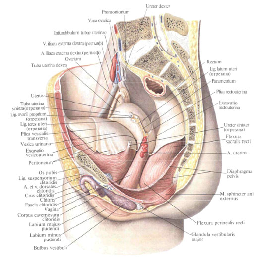
The vagina , vagina, is a tubular, flattened anteroposterior organ. The length of the vagina is 8-10 cm. Its upper border is located at the level of the cervix, which it covers, at the bottom it opens on the threshold of the vagina with a vaginal opening , ostium vaginae. The vagina is located from top to bottom and from behind in front, corresponding to the axis of the lower segment of the small pelvis; Relative to the uterus, the vagina forms an angle open anteriorly. The front wall, paries anterior, and posterior wall, paries posterior, the vagina touch, resulting in the vaginal cavity has a slit shape. The anterior wall of the vagina due to the fact that it connects the tissue of the urethra, thicker than the posterior. 
At the very top, the vaginal cavity forms a blind pocket - the vaginal fornix , fornix vaginae - around the protruding cervix of the uterus, in which the fore part, pars anterior, posterior part, pars posterior, and lateral part, pars lateralis, are distinguished. The part that lies between the posterior lip of the cervix and the back wall of the vagina is deeper than the section between the anterior lip and the anterior wall of the vagina. The surrounding tissue of the vagina connective tissue with an admixture of a small number of smooth muscle fibers is particularly dense in the lower parts, where the vaginal wall joins the rectum, bladder and urethra.
The structure of the vagina, function, the walls of the vagina.
The walls of the vagina consist of three layers: muscular, mucous and underdeveloped spongy membranes.

The muscular membrane , tunica muscularis, consists of two layers of muscles: external longitudinal and internal circular. In the region of the pelvic diaphragm, the fibers of both layers are partly intertwined. Here, from the muscles of the pelvic floor, longitudinal transverse striated muscle bundles join. In the upper part of the vagina there are only smooth muscle fibers.
The mucous membrane, tunica mucosa, is tightly fused to the muscular not all over. It is much thicker than the muscular, in some areas its thickness reaches 2 mm. On its walls, especially in the lower part, there are transverse folds - vaginal folds, rugae vaginales. In the middle sections of the anterior and posterior walls of the vagina, the transverse folds protrude more than on the sides, forming here longitudinal elevations - columns of folds, columnae rugarum. Distinguish between the anterior and posterior columns of folds, columnae rugarum anterior and posterior. The lower end of the anterior post is called the urethral keel of the vagina, carina urethralis vaginae, since it is subject to the lower part of the urethra.
All these folds cause significant extensibility of the mucosa, and with it all layers of the vaginal walls, which better ensures the passage of the fetus through the birth canal. In the area of vaginal folds and pillars of folds, between the mucous membrane and the muscular membranes, a thin spongy shell is deposited.
The walls of the vagina adhere to the organs of the cavity of the small pelvis. The front wall is loosely fused with the bladder and a dense connective tissue - with the urethra. In the anterolateral areas of the vagina is the ureter. The back wall, with the exception of the upper portion covered by the peritoneum (about 1/4 of the wall), is fused to the anterior wall of the rectum in the region of the ampulla of the gut. The posterior wall in its middle sections covers the bundles of muscle that lifts the anus.
In women who did not live sexually, in the area of the vaginal opening on the back and partly lateral edges there is a thin fold of the mucous membrane - the hymen, hymen, usually a semilunar one. After its rupture, sometimes there are so-called hymen flaps, carunculae hymenales, irregularly shaped, having the appearance of papillae. The location of these papillae is considered the lower border of the vagina.
The lower part of the vagina passes through the urogenital diaphragm.
Innervation: plexus hypogastricus inferior (sympathetic innervation), nn. Splanchnici pelvici (parasympathetic innervation), n.pudendus (the lower part of the vagina).
Blood supply: aa. Vaginales (from A. uterina), a. Rectalis media, a. Vesicalis inferior (a. Pudenda interna). Venous outflow is carried out in plexus venosus vaginalis and then to v. Iliaca interna. Lymphatic vessels direct lymph to the nodi lymphatici inguinalis interni (from the upper part) and to the nodi lymphatici inguinalis (from the bottom).

Comments
When commenting on, remember that the content and tone of your message can hurt the feelings of real people, show respect and tolerance to your interlocutors even if you do not share their opinion, your behavior in the conditions of freedom of expression and anonymity provided by the Internet, changes Not only virtual, but also the real world. All comments are hidden from the index, spam is controlled.