


Development and age specific features of the urogenital organs
Development and age specific features of the urogenital apparatus.
The organs of the urinary system to the reproductive system, in spite of various functional features, are similar in development. In the formation of the organs of these systems, the mesoblast is involved, from which the kidneys and genital glands are formed, and the ectoblast lining the cloacal cavity and the enteroblast of the posterior part of the intestinal tube participate in the formation of the excretory urinary and genital tract and reproductive organs. The organs of the urinary system - the kidneys - are laid somewhat before the genital organs and pass through three stages in their development: pronephros, primary mesonephros and secondary or metanephros.
Preferences are laid in the middle of the third week, the primary kidney is in the middle of the 4th week of the intrauterine period and is rapidly reduced, taking part in the development of male genital tracts. Both stages still in the intrauterine period are replaced by the stage of the final kidney, which persists for life.
In the kidneys of the newborn lobularity is clearly expressed (on average 14 lobules), which is caused by the insufficient development of cortical substance. Lobble disappears to 2-4 years of life.

The length of the kidney in a newborn is 3.5-4.0 cm, width 1.7-2.1 cm, thickness 1.6 cm; The mass of the kidney is 11-12 g. By the beginning of the second year of life, the size of the kidney is almost doubled. In the newborn, the upper pole of the kidney lies at the level of the lower edge of the body of the XI thoracic vertebra; In 3-5 months - at the level of the middle of the XII thoracic vertebra; To 2 years of age reaches the level of the kidney of an adult. The lower pole of the right kidney is located at the level of the lower edge IV of the lumbar vertebra, the left kidney is at the level of the middle of the body of the IV lumbar vertebra. Depending on the location of the kidneys, the renal arteries and veins are obliquely positioned relative to the exit from the aorta or into the lower vena cava.
The kidneys of the newborn in their position correspond to the level of the II lumbar vertebra, and in the adult - to the level I of the lumbar vertebra.
On the incision of the newborn's kidney, an underdeveloped cortical substance is visible, in which the convoluted tubules are not sufficiently developed. The cortical substance by 9-10 years becomes the same as the cortical substance of the kidney of an adult. The brain substance develops more intensively; The ratio of cortical and cerebral matter in a newborn is 1: 4, in an adult 1: 2. The kidneys of the newborn are covered with their own fine fibrous capsule, which is well developed by the age of 6-7.
The renal pelvis and the uterus of the newborn have some characteristics. The pelvis is relatively broader and the ureters have a more convoluted direction than the adult.
There are indications that if the lobe of the kidney is well expressed, then the pelvis and ureter are wider.
The bladder develops from the so-called endodermal rudiment, which is formed by joining the ventral section of the cloaca with the allantois. The newborn's bladder is fusiform, its upper portion narrowed. This form remains up to 1.5 years, up to 5 years it resembles a plum, and by the age of 10 it becomes ovoid. At the age of 15-17 takes the form of an adult's bladder. The internal opening of the urethra in the newborn is often located at the level of the upper edge of the symphysis.
The curvature of the male urethra of a newborn resembles that of an adult. The length of the newborn urethra reaches 5-5.5 cm, by the age of 10 it is doubled, and by the age of 16 it is 16-17 cm, and the greatest growth is observed at the age of 13-14. In newborn girls, the length of the urethra is about 1 cm, and by the age of 16 it reaches 3.5-4.0 cm. The inner opening of the urethra lies at the level of the middle of the pubic symphysis, and the urethra itself lies on the posterior surface of the urethra, but as the pelvic floor descends, And with it the bladder it straightens.
Male gland - testicle - is laid in the abdominal cavity and on the 11-12th week approaches the deep inguinal ring. Then, starting from the 24-25th week, gradually through the inguinal canal the testicle descends into the scrotum. This process ends at the time of birth. Together with the testicle, the appendage and part of the vas deferens down into the scrotum.
The prostate gland is formed from the urethral epithelium by the end of the 3rd month of the intrauterine period. The prostate gland is developing very slowly. It increases slightly to the 6th to 10th year of life, reaching considerable dimensions during puberty. In the newborn it is spherical, with age it becomes somewhat flattened, and by 15-16 years has a heart shape. Finally, the gland develops to 17 years.
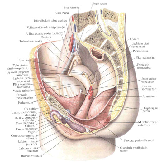
Uterus , fallopian tubes , vagina develop from parameconephalic ducts, which, going to the cavity of the future pelvis, undergo great changes; Their upper sections form the fallopian tubes, the middle and lower divisions merge between the right and left ducts and form the uterus, the vagina is formed from the lower parts, and the mesentery of the primary kidneys form the broad ligament of the uterus.
In the newborn, the uterus has a length of up to 3.5-4.0 cm, its mass is 2 g. Shortly after birth, there is an involution: the length of the uterus reaches 2.5 cm. In early childhood, the uterus has an elongated shape and is somewhat compressed from front to back. By 8-9 years the uterus body takes a round shape; By the age of 12-14, the uterus acquires a pear-shaped form and soon becomes a uterus of an adult woman. The vagina of a newborn has a length of up to 3 cm. Its position depends on the gradual lowering of both its and the bladder: their topografical and anatomical relationship is changing. In early childhood, the vagina with the uterus forms an obtuse angle; Its front wall is somewhat shorter than the rear. Fallopian tubes of the newborn are convoluted, their free ends are located farther from the ovaries than in the adult woman. Since the age of five, the fallopian tubes and ovaries are located in the same way as in an adult woman.
In newborns, the ovaries lie at the level of the cape or somewhat lower in the region of the large pelvis and only 5 years fall into the small pelvis, occupying a position that is typical for an adult woman. The ovary of the newborn is an elongated flat organ, which becomes ellipsoidal by the age of 10. The surface of the ovary in the early childhood is smooth, and with age, scars appear on it.
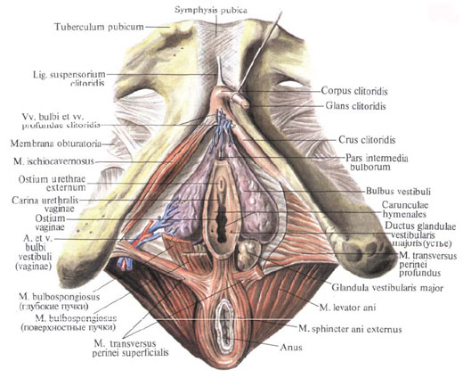
The development of the external genitalia begins on the 8th-9th week, from the moment of the formation of the sexual tubercle, the genital folds and the surrounding sexual roller. In men from the genital tubercle and genital folds, the penis develops, and the genital shaft forms the scrotum. In women, the genital tubercle develops into the clitoris, the sexual folds into the small labia, and the genital cushion into the large labia.

Comments
Commenting on, remember that the content and tone of your message can hurt the feelings of real people, show respect and tolerance to your interlocutors even if you do not share their opinion, your behavior in the conditions of freedom of expression and anonymity provided by the Internet, changes Not only virtual, but also the real world. All comments are hidden from the index, spam is controlled.