The adrenal glands. Structure, functions
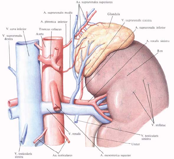
Adrenals, glandulae suprarenales (adrenales), paired, each of them is located at the level of XI and XII thoracic vertebrae above the kidney , on the upper medial part of its upper end. Adrenals lie in the retroperitoneal cellulose and are embedded in the renal fascia.
The right adrenal triangle, already located above the left, lies above the upper pole of the right kidney, immediately adjacent to the inferior vena cava. At its widest length, it is covered by the peritoneum, with the exception of the lower part of the anterior surface, to which it adjoins the liver , leaving the impression on the latter, impressio suprarenalis.
The left adrenal gland is semilunar, located partly above the upper pole of the left kidney and partially adjacent to its medial margin. It is covered with the peritoneum in front, mainly in its upper part. The left adrenal gland is in contact with the cardiac part of the stomach, the spleen and the pancreas . Both adrenal glands adjoin the diaphragm behind.
 The structure of the adrenal glands
In each adrenal gland the front surface is distinguished, the facies anterior, posterior surface, posterior facies, and a concave shape of the renal surface, the facies renalis, which adrenal adheres to the corresponding kidney. In addition, the upper margin, margo superior, and the medial margin, margo medialis, are distinguished.
The anterior and posterior surfaces of the adrenal gland are covered with furrows. The deepest furrow, located on the anterior medial surface, received the name of the gate, hilum.
In the right adrenal gland the gates lie closer to the top of the gland, in the left - closer to the base. A central vein emerges through the gate, v. Centralis, which on exit is called the adrenal vein, v. Suprarenalis. The last of the right gland runs into the inferior hollow vein, from the left - into the left renal vein. In the gates lie and lymphatic vessels of the adrenal gland, while the arterial branches and nerve trunks can penetrate into the thickness of the gland from the front and back surfaces.
The mass and dimensions of the adrenal gland are individual. Thus, the mass of each gland varies from 11 to 18 g in an adult (or from 7 to 20 g), in a newborn is 6 g. The longitudinal size is up to 6 cm, the transverse dimension is up to 3 cm, and 1 cm thick (sometimes larger).
Outside, the adrenal gland is covered with a thin fibrous capsule with an admixture of smooth muscle fibers; From the capsule, the processes extend into the thickness of the gland.
Parenchyma of the adrenal gland consists of two layers - the outer cortex (cortex), cortex, and the inner medulla, medulla, differing in development and in function.
The outer layer is thicker, yellowish brown, formed by glandular and connective tissue. The inner layer is brownish red, contains chromaffin and sympathetic nerve cells.
Occasionally there are additional adrenal glands , glandulae suprarenales ascessoriae, which can be a cortical or brain substance lying in the retroperitoneal tissue.
The cortex of the adrenals produces a large number of hormones - corticosteroids, which include three main groups: mineralocorticoids (aldosterone), glucocorticoids (hydrocortisone, corticosterone) and sex hormones (androgens). The action of these hormones is very diverse. They enhance the reabsorption of sodium, promote the release of potassium ions and the concentration of chlorine in the blood, and also participate in the regulation of body metabolism: carbohydrate, fat, protein and water-salt.
Hormones of the medulla are adrenaline and norepinephrine , which enhance the excitation and contraction of the heart muscle. Simultaneously, hormones increase the tone of the sympathetic part of the autonomic nervous system, providing a vasoconstrictor effect, which causes an increase in blood pressure.
Innervation: branches from plexus celiacus, renalis, suprarenalis, which contain sympathetic fibers and fibers of the wandering and diaphragmatic nerves.
Blood supply: a. Suprarenalis superior (from a. Phrenica inferior), a. Suprarenalis media (from aorta abdominalis), a. Suprarenalis inferior (from A. renalis), their branches under the capsule of the adrenal gland form a vascular arterial network, the trunks of which penetrate into the gland. Venous blood flows out by v. Centralis, located intraorganically, in v. Suprarenalis (empties to the right in v. Cava inferior, left - in v. Renalis sinistra). Lymphatic vessels flow into the nodi lymphatici lumbales that surround the aorta and inferior vena cava.


Comments
When commenting on, remember that the content and tone of your message can hurt the feelings of real people, show respect and tolerance to your interlocutors even if you do not share their opinion, your behavior in the conditions of freedom of expression and anonymity provided by the Internet, changes Not only virtual, but also the real world. All comments are hidden from the index, spam is controlled.