


Paraorthal bodies
PARAORTHAL TELTS.
In the course of the aorta there is a significant amount of paraganglia - paraaortic bodies , corpora paraaortica, which can be topographically divided into a number of groups.
These include: 1) intact paragangia, 2) aortic glomus, and 3) coccygeal body.
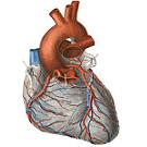
1. Non-cardiac paraganglia - formation in the form of two clusters - upper and lower, paraganglion supracardiale, superius et inferius. Each of them is unstable, especially the lower, which decreases with age. The upper cluster in an adult is larger in size than the lower one; It is located between the pulmonary trunk and the aorta, in the region of its arc, being tightly joined to it; The lower one lies at the exit site of the left coronary artery. The vertex paragangia is associated with the nerve plexus located at the site of its bedding.
In the thickness of the paraganglia lies a multitude of blood vessels.
In addition to these paraganglia, there are separate subpericardial accumulations of chromaffin cells.

2. Aortic glomus, glomera aortica, is divided into the right and left parts. Each part is a stretch along the lateral surface of the abdominal aorta, the right is slightly longer than the left (the length of the right oscillates from 8 to 20 mm, the left - from 8 to 15 mm, thickness 2-3 mm). Both strands are sometimes interconnected by a small, almost transversely extending strip of chromaffin tissue located along the anterior surface of the aorta. Aortic paraganglia are associated with the branches of the lower thoracic and lumbar nodes of the sympathetic trunk.
The blood supply of paraganglia is provided by the nearest vessels located in the surrounding cellulose (branches of the abdominal aorta, intestinal arteries, etc.). Venous blood is collected by the lower hollow and left renal veins.
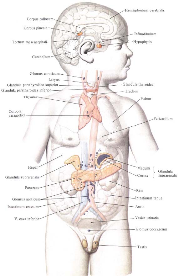
3. Coccygeal glomus, glomus coccygeum, is an unpaired formation up to 2.5 mm long, located on the front surface of the tip of the coccyx.
The blood supply of the coccygeal glomus is associated with the thin branches that extend from the middle median sacral artery. In the thickness of the organ itself, the vascular network branches out into glomeruli.
In addition to the listed large accumulations of chromaffin tissue, in fetuses and children of early age, other accumulations of this tissue scattered in various amounts are encountered in places genetically related to the autonomic nervous system, its sympathetic part; In a number of sites of retroperitoneal tissue of the lumbar aorta, in the ovaries, testes, along the wall of some vessels, etc.


Comments
When commenting on, remember that the content and tone of your message can hurt the feelings of real people, show respect and tolerance to your interlocutors even if you do not share their opinion, your behavior in the conditions of freedom of expression and anonymity provided by the Internet, changes Not only virtual, but also the real world. All comments are hidden from the index, spam is controlled.