


Language
The tongue , lingua, is the muscular organ, covered from above, from the sides and partly from below by the mucous membrane. In the language, two parts are distinguished: the front, free, part, or body of the tongue, corpus linguae, and the back is the root of the tongue, radix linguae.
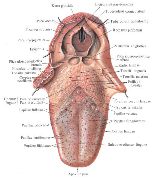
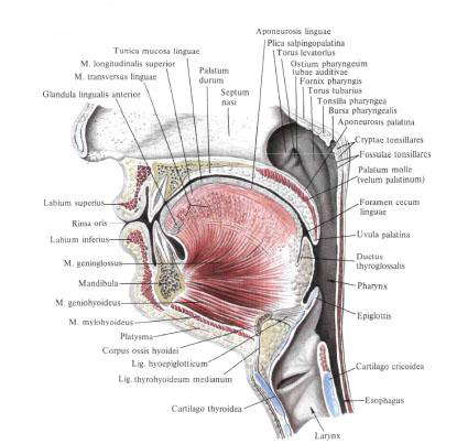
The body of the tongue , corpus linguae, ends at the front with a flat, rounded apex of the tongue, apex linguae; Back, the body is delimited from the root by the boundary furrow, sulcus terminalis, hence the names: the pre-species, pars presulcalis, and the post-partum part, pars postsulcalis, lies beyond the border groove and is the root of the tongue, radix linguae. The borderline furrow consists of two halves that converge on the median line of the tongue at an obtuse angle open anteriorly. At the top of this obtuse angle there is a blind hole of the tongue, foramen cecum linguae, - the trace of the overgrown tongue-tongued duct, ductus thyroglossalis.
Upper, rear, surface - back of tongue, dorsum linguae, - convex in longitudinal and transverse directions; On it in the longitudinal direction is located the median groove of the tongue, sulcus medianus linguae, which divides the body of the tongue into the right and left parts. Accordingly, this groove in the thickness of the tongue is a connective tissue plate - the septum of the tongue, septum linguae. The body of the tongue is bounded on the sides by the edge of the tongue, margo linguae.
The lower surface of the tongue, the facies inferior linguae, is free only in its anterior part. Its mucosa is smooth and has two frontal frontal folds, plicae fimbriatae. From the lower surface of the tongue to the gums in the sagittal direction there is a fold of the mucous membrane - the frenulum of the tongue, frenulum linguae.
Muscles of the tongue.
The tongue is the muscular organ. The muscles of the tongue, mm. Linguae, can be divided into two groups. One is represented by muscles starting on the bones and weaving into the body of the tongue. These muscles are called skeletal muscles, their contraction changes the position of the tongue. Another group of muscles is the language's own muscles, their function is to change the form of the language.
Skeletal muscles of the tongue
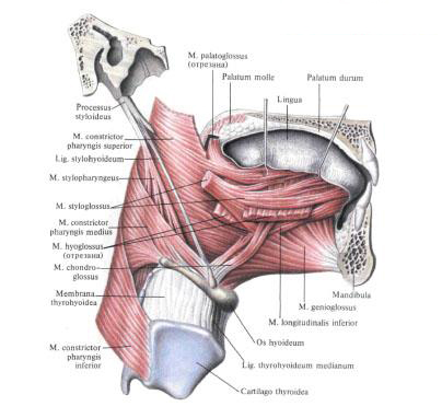
- Stylococcus aureus, m. Styloglossus, starts from the styloid process and the prosthetic ligament, goes obliquely downward, forward and inward, between m. Stylohyoideus and pharynx , is attached to the lateral surface of the root of the tongue and the outer surface of the hyoid-lingual muscle. A thicker upper beam of it is directed along the edge of the tongue to its apex; The thinner lower fascicle perforates the sublingual-lingual muscle and is guided inwards at the back of the tongue, where it is weaved by tendon bundles with the same-named muscle of the opposite side. Function: pulls the tongue, especially the root of it, up and back.
- Sublingual-lingual muscle, m. Hyoglossus, flat, quadrangular, lying outside of the chin-lingual muscle. It starts from the upper edge of the body and the big horn of the hyoid bone . Its tufts are directed upward and anteriorly, to the lateral edge of the root and body of the tongue, where, passing between m. Styloglossus and m. Longitudinalis inferior, reach the tip of the tongue. Function: pulls the tongue back and down.
- The chin-lingual muscle, m. Genioglossus, is located on the sides of the septum of the tongue. It starts from the chin awn, from which its tufts, fan-likely diverging, follow the mucous membrane of the tongue throughout its entire length. The lower muscle tufts running above m. Geniohyoideus, are attached to the body of the hyoid bone and epiglottis. Function: pulls the language forward and down.
- Cartilaginous muscle, m. Chondroglossus, in the form of a small muscle bundle begins on the small horn of the hyoid bone and weaves into the thickness of the mouse's tongue in the area of its back. Function: pulls the tongue back and down.
Own muscles of the tongue
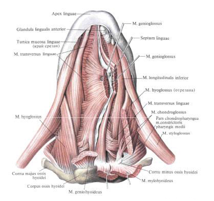
- Lower longitudinal muscle, m. Longitudinalis inferior, long and narrow, lies and thicker than the tongue, outside of m. Genioglossus. It starts from the aponeurosis of the tongue, in the region of the root of the tongue, and goes to the tip of the tongue, ending on its lower surface. The initial parts of the muscle are located between m. Hyoglossus and m. Genioglossus, and then between m. Styloglossus, m. Genioglosus. Function: shortens the language.
- Upper longitudinal muscle, m. Longitudinalis superior, originates in three bundles: medial - from the anterior surface of the epiglottis and from the middle lingual-epiglottis fold and two lateral - from the small horn of the hyoid bone . All three bundles then converge and go directly under the aponeurosis of the tongue and mucous membrane, along the entire back of the tongue to its apex; While all along the way the bundles are intertwined. Function: participates in the bending of the tongue, shortens the tongue and lifts its tip.
- The transverse muscle of the tongue, m. Transversa linguae, lies all over the tongue. It consists of individual transversely moving muscle beams, starting from the septum of the tongue, partly perforating it; Ends in the aponeurosis and mucosa of the edges and back of the tongue. Function: reduces the transverse diameter of the tongue and makes it transversely convex upward.
- Vertical muscle of the tongue, m. Verticalis linguae, consists of short beams located in the free part of the tongue between its back and the lower surface. Function: flatten the language.
Innervation: all muscles of the tongue - rr. Linguales n. Hypoglossi.
Blood supply: all muscles of the tongue - a. Lingualis . Venous blood flows out by v. Lingualis, which flows into v. Jugularis interna.
The lymph flows to the nodi lymphatici submandibulares and cervicales laterales profundi.
Mucous membrane of the tongue.
The mucous membrane of the tongue, tunica mucosa linguae, is smooth in the root region, the lower surface of the body and the apex and is roughen on the back of the tongue. This roughness is caused by a large number of small elevations - papillae of the tongue, papillae linguales. All papillae of the tongue differ in shape.
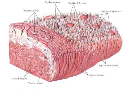
- Filiform papillae, papillae filiformes, are located on the entire body of the tongue, impart a velvety texture to the mucous membrane. They are formations consisting of a projection of the mucous membrane with racemose epithelial appendages on the apexes. Filiform papillae are most pronounced in the middle section of the back of the tongue and close to the tubal papillae.
- Cone-shaped papillae, papilae conicae, less filamentary in size, but their conical, horny apexes protrude somewhat above the surface of the mucous membrane. They are located in the central sections of the back of the tongue, closer to the boundary groove.
- Mushroom papillae, papillae fungiformes, number from 150 to 200, are scattered along the back of the tongue, closer to its edges, especially in the apex region, are rare in the middle section of the tongue. They are in the form of mushroom-shaped outgrowths, larger than filamentous and therefore well distinguishable between them.
- Lenticular papillae, papillae lentiformes, lie along the edges of the tongue among mushroom papillae. There are large ones among them, the apices of which are considerably flattened, and the papillae below the mushroom ones.
- Tubular papillae, papillae vallatae, number from 7 to 11, are the largest, but they are not very prominent above the surface; Are located on the boundary between the body and the root of the tongue, anterior to the boundary furrow and parallel to it. The central papilla, surrounded by a shaft, lies in front of the blind opening of the tongue. Each papilla consists of a small cylindrical elevation surrounded by an annular groove, around which there is a roller of the mucous membrane.
- The leaf papillae, papillae foliatae, are located on the lateral parts of the tongue. They consist of 5-8 furrows separated by grooves, extending almost vertically in front of the tongue-and-tongue arch. The leaf papillae are not the same in size and are better expressed nearer to the root of the tongue.
On the mucous membrane of the tongue open the ducts of many lingual glands, glandulae linguales, belonging to the small salivary glands. In addition, under the epithelium in the area of the root of the tongue to the epiglottis lies a large number of lymphatic follicles, folliculi limphatici, of various sizes.
All the accumulation of lingual follicles, folliculi linguales, was called lingual tonsils, tonsilla lingualis.
The lingual glands , glandulae linguales, are grouped into mucous, serous and mixed glands. Among them are several glands.
Anterior lingual gland, glandula lingualis anterior, in the form of oblong formation is located on both sides of the anterior end of m. Genioglossi, near and behind the tip of the tongue. The outlet ducts open on the lower surface of the tongue along the fringe fold. In addition, these glands can be located in the form of small groups along the edges of the back of the body of the tongue at the back of the muscles (mm. Styloglossus and palatoglossus). The opening ducts open them in the folds of leaf-shaped papillae.
The glands of the lingual tonsil region form under the mucous membrane a layer 4-8 mm thick, occupying the area of the lingual tonsil to the epiglottis. Their excretory ducts open into the grooves located around the follicles, and into the indentation in the middle of the follicle.
The glands of the region of the trough-shaped and leaf-shaped papillae are serous glands. Their ducts open into the grooves surrounding the rachetical papillae, and into the folds between the papillae.
The mucous membrane, moving from the root of the tongue to the epiglottis, forms three folds. One of them, unpaired, is the central-middle lingual-epiglottis fold, plica glossoepiglottica mediana; The paired fold goes to the lateral edge of the epiglottis - lateral lingual-epiglottis fold, plica glossoepiglottica lateralis. Between the middle and lateral folds on each side is the fovea of the epiglottis, vallecula epiglottica.
In the submucosal base of the tongue lies a large amount of loose connective tissue and tendon pinches of the own muscles of the tongue, which together form a powerful aponeurosis of the tongue, aponeurosis linguae, to which the muscles of the tongue are attached.
Vessels and nerves pass through the tongue.
Innervation: the front two thirds - n. Lingualis, chorda tympani; The back third is nn. Glossopharyngeus, laryngeus superior.
Blood supply: a. Lingualis . Venous blood flows out by v. Lingualis, which flows into v. Jugularis interna. The lymph flows to the nodi lymphatici submentales, submandibulares and cervicales laterales profundi.
Mucous membrane of the bottom of the oral cavity.
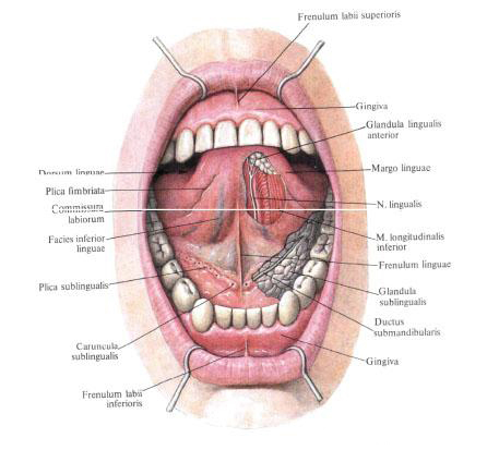
At the bottom of the oral cavity, on either side of the tongue bridle, lies a small round tubercle - the sublingual papilla, caruncula sublingualis, in which the ducts of the submandibular and hyoidal glands open: the submandibular duct, ductus submandibularis, and the large hyoid duct, ductus sublingualis major.
Behind and outside of the hyoid papilla mucosa covers the hyoid gland, forming a longitudinally extending hyoid fold, plica sublingualis; On this fold small sublingual ducts, ductus sublinguales minores, are opened.
You will be interested to read this:

Comments
When commenting on, remember that the content and tone of your message can hurt the feelings of real people, show respect and tolerance to your interlocutors even if you do not share their opinion, your behavior in the conditions of freedom of expression and anonymity provided by the Internet, changes Not only virtual, but also the real world. All comments are hidden from the index, spam is controlled.