


Esophagus, function, structure of esophagus
Esophagus, esophagus, looks like a tube that connects the pharynx with the stomach . The place of passage of the pharynx into the esophagus in an adult corresponds to the level of the sixth cervical vertebra or the lower edge of the cricoid cartilage, and the place of passage into the stomach is projected at the level of the XI thoracic vertebra . In a living person, these boundaries can change when you roll your head, take a deep breath or lower your stomach. The length of the esophagus is up to 25 cm. 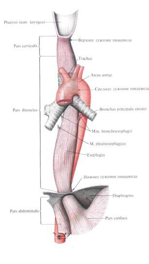
A small part of the esophagus lies in the neck, then the esophagus passes through the upper aperture of the thorax into the chest cavity, and then passes through the esophagus through the esophageal aperture of the diaphragm into the abdominal cavity, passing into the cardiac part of the stomach. In this connection, three parts are distinguished in the esophagus; Neck part, pars cervical is, thoracic part, pars thoracica, and abdominal part, pars abdominalis.
Cervical honor, pars cervicalis, is located from the level of the sixth cervical vertebra to the I-II thoracic. Its length varies from 5 to 8 cm.
The thoracic part, pars thoracica, has the largest length - 15-18 cm and ends at the level of IX-X thoracic vertebrae, i.e. At the place of entry of the esophagus into the esophageal opening of the diaphragm.
Abdominal part, pars abdominalis. The shortest, its length is 1-3 cm.
The esophagus lies in front of the vertebral column and has 4 bends in its path: two in the sagittal plane and two in the frontal plane.
The initial section of the esophagus is located almost strictly on the median line. At level II of the thoracic vertebra, the esophagus deviates to the left, occupying the region III And IV Vertebrae leftmost position. Then, at level V of the vertebra, it again lies on the median line, and below it comes somewhat to the right of it. The bend to the right extends to VIII Thoracic vertebra. Driving downwards, the esophagus is at the level of VIII Up to the X vertebra again goes to the left side. These two bends lie in the frontal plane. The first bend in the sagittal plane of the esophagus under the bifurcation of the trachea - here it deviates posteriorly. The second bend in this plane is marked on the level of VIII-IX vertebrae, corresponding to the place of passage of the esophagus through the diaphragm - here the esophagus deviates anteriorly.
In its turn, the esophagus adjoins a number of organs.
The neck part of the esophagus with its posterior surface lies on the prevertebral plate, and the anterior surface adjoins the membranous wall of the trachea. From the sides to the esophagus, common carotid arteries and recurrent laryngeal nerves are close in this department.
The thoracic part of the esophagus with its posterior surface also lies along the spine, and the upper third of the anterior surface adjoins the membranous wall of the trachea. Then, at the level of IV-V of the vertebrae, the esophagus is attached to the aortic arch by the anterior surface, and below the latter, it adjoins the posterior surface of the left bronchus, connecting with it with the aid of an underdeveloped bronchopiscusis muscle, m. Bronchoesophageus. The musculature is paired, unstable, is a muscular-elastic stretch attached to the posterior surface of the main bronchus.
In the lower third, the esophagus touches the pericardium corresponding to the left atrium and left ventricle, and, going down, spirals around the aorta, passing into the abdominal part. The latter is covered at the front with a portion of the left lobe of the liver. Along the lower part of the thoracic part of the esophagus, a rear wandering X-pair trunk is attached to its rear surface, and the front wandering trunk is to the front.
The lumen of the esophagus is not the same. Throughout its length it is customary to distinguish three constrictions and two extensions. The first constriction is located at the site of the pharyngeal transition into the esophagus, the second is where the esophagus is adjacent to the arch of the aorta, and the third is at the site of passage through the esophageal opening of the diaphragm. Between these restrictions there are two extensions.
The wall of the esophagus has three membranes: mucous, muscular and adventitial; The ventral part is covered with a serous membrane.
The mucous membrane, tunica mucosa, is covered with multilayered flat epithelium. The thickness of the mucous layer is formed by loose fiber and a developed muscular plate of the mucous membrane, lamina muscularis mucosae, consisting of smooth fibers, whose role reduces to contracting the mucous membrane with a decrease in the lumen of the esophagus.
In the transverse section, the lumen of the esophagus has the appearance of a stellate slit due to compressed walls and well-defined longitudinal folds. The size of the folds is due to the considerable development of the loose connective tissue that forms the submucosa, tela submucosa. The latter lies between the mucosa and the muscular membranes. In the submucosa base there are many vessels, esophagus glands, glandulae esophageae, the ducts of which open on the surface of the mucous membrane, and single lymphatic follicles.
The muscular membrane, tunica muscularis, consists of two layers: the inner - the circular and the outer - the longitudinal,
In the intermuscular layer, in its loose connective tissue, there are vascular networks and neural plexuses,
In the upper third of the esophagus, the muscle layers are represented by striated muscle, which in the middle third passes into a smooth one; The lower third of the esophagus consists exclusively of smooth muscle fibers. Muscular layers are unevenly developed. Thus, the longitudinal layer is composed of fibers that separate in the upper part of the esophagus in the ring-esophageal tendon, tendo cricoesopha-geus, a pair that attaches to the lower edge of the plate of the cricoid larynx of the larynx. Therefore, in the initial part of the esophagus there is a site without a longitudinal elephant. The circular layer of the esophagus wall in the upper regions is a continuation of the pharyngeal musculature, and below it passes into circular and oblique fibers of the muscular wall of the stomach. In some parts of the esophagus it is possible to see an underdeveloped longitudinal layer, which lies inward from the circular one.
At the level of the collar of the lungs from the esophagus, the paired pleurosophageal muscle departs, m. Pleuroesophageus, consisting mainly of smooth muscle fibers. The left muscle connects the aorta and esophagus with the mediastinal pleura at the level of the bifurcation of the bronchi, and to the right extends from the lower third of the thoracic part of the esophagus and approaches the right mediastinal pleura.
The adventitia, tunica adventitia, is formed by a loose connective tissue containing a small amount of elastic fibers. With the help of this shell, the esophagus is fixed to other organs that surround it in the posterior mediastinum. In the thickness of this shell pass the main blood vessels that carry blood supply to the esophagus, lymph vessels carrying lymph from the walls of the esophagus, and also the nerve trunks of the vagus nerves, which form plexuses here.
Innervation: plexus esophageus (n. Vagus and truncus sympathicus) is the source of the powerful intra-wall plexuses of the nitegu.
Blood supply: neck part - rr. Esophageales from a. Thyreoidea inferior; Thoracic part - rr. Esophageales or aorta thoracica, abdominal part - rr. Esophageales from a. Gastrica sinistra and a. Phrenica inferior sinistra. Venous blood flows from the cervical part in v. Thyreoidea inferior, and then v. Brachiocephalica; From the thoracic part to v. Azygos and v. Hemiazygos: from the ventral part to v. Gastrica sinistra, and then v. Portae. Lymph flows from the cervical part in the nodi lymphatici tracheobronchiales superiores et inferiores, paratracheales and paraverlebrales: from the thoracic part in the nodi lymphatici tracheobronchiales inferiores and mediastinals posteriores: from the ventral part to the anulus lymphatici cardii.
You will be interested to read this:

Comments
When commenting on, remember that the content and tone of your message can hurt the feelings of real people, show respect and tolerance to your interlocutors even if you do not share their opinion, your behavior in the conditions of freedom of expression and anonymity provided by the Internet, changes Not only virtual, but also the real world. All comments are hidden from the index, spam is controlled.