


Rectum, structure, functions
The rectum , rectum, is located in the cavity of the small pelvis, at the back of its wall, formed by the sacrum, coccyx and the back of the muscles of the pelvic floor. It starts from the end of the pelvic part of the sigmoid colon at level III of the sacral vertebra and ends in the crotch area in the anus. Its length is 14-18 cm. The diameter of the rectum varies from 4 cm (the beginning of the sigmoid colon) to 7.5 cm in the middle part (ampulla), and again decreases to a gap at the level of the anus.
The rectum consists of two parts: pelvic and perineal. The first is located above the diaphragm of the pelvis, in the cavity of the small pelvis, and in turn is subdivided into a narrower nascent pulp and a large ampulla of the rectum, ampulla recti. The second part of the rectum lies under the pelvic diaphragm, in the perineal region, and represents the anal (anal) canal, canalis analis.
The pelvic part of the rectum forms in the sagittal plane a bend open anteriorly to the concavity of the sacrum, a sacral bend, flexura sacralis; The upper part of the gut bend is from front to back and down, lower - from behind in front and down.
In the frontal plane, the pelvic part forms unstable bends; The upper part of the bend goes to the left from top to bottom and to the right, the bottom - in the opposite direction. The second bending in the sagittal plane, but already concave back, is located at the transition of the pelvic part into the perineal; After passing the diaphragm of the pelvis, the rectum turns sharply (almost at right angles) back, forming a perineal flexure, flexura perinealis. At this level, the rectum as it passes around the tip of the tailbone. The length of the pelvic part varies from 10 to 14 cm, the crotch part is about 4 cm.
At the level of the lower edge of the III sacral vertebra, the rectum begins to lose its serous cover: first from the posterior surface, then from the lateral and, finally, from the anterior. Thus, the upper, namdapular, part of the pelvic part of the rectum is located intraperitoneally, the upper part of the ampulla is surrounded by the serosa from three sides, and the lowest part of the ampulla lies retroperitatively, since the peritoneum covers here only a small portion of the anterior wall.
The line along which the peritoneum leaves the wall of the intestine follows the obliquely from above, from behind downwards and forward. As the pelvic wall of the rectum loses its peritoneal cover, it is replaced by the visceral fascia of the pelvis, which forms the case of the rectum.
The crotch part of the rectum looks like a longitudinal slit and opens in the deepening of the interannual sulcus in the anus, anus, almost in the middle of the distance between the coccyx and the scrotum root in men or the posterior adhesion of the large labia in women, at the level of the transverse line connecting both sciatic tubercles.
The structure of the rectum wall.

Serous membrane (peritoneum), tunica serosa, is a part of the rectum wall only on a small extent. The extraperitoneal part of the pelvic part of the rectum is surrounded by the visceral fascia of the pelvis, the fascia is not directly attached to the muscular layer of the intestinal wall. Between the visceral fascia and the muscular layer lies a layer of fatty tissue, nerves are located, which feed the intestine blood vessels and lymph nodes. The anterior section of the fascia of the rectum is a plate that separates the bowel from the organs lying ahead: the bladder, prostate, etc. This plate is a derivative of the fused serous sheets of the deepest part of the peritoneal pocket of the small pelvis; It goes from the bottom of the rectum-uterine cavity (or rectal-vesicular cavity in men) to the tendon center of the perineal muscles and is called the peritoneal-perineal fascia, fascia peritoneoperinealis, or rectilocephalic septum, septum rectovesicale. Dorsal rectum fascia ends in the middle line of the posterior wall of the rectum.
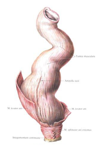
The muscular membrane, tunica muscularis, of the rectum consists of two layers: the outer longitudinal, stratum longitudinale, less thick, and the inner circular, stratum circulare, thicker. The longitudinal layer is an extension of the sigmoid colon muscle bands, which expand here and cover the gut with a continuous layer. On the anterior and posterior walls the longitudinal muscle bundles are more developed. In the longitudinal muscular layer of the lower part of the ampoule, the bundles coming from the anterior sacrococcygeal ligament are interlaced, the rectum-coccygeal muscle, m. Rectococcygeus. A part of the muscle fibers of the longitudinal layer interweaves in the muscle lifting the anus, m. Levator ani, and the part reaches the skin of the anus.
In men on the anterior surface of the lower part of the rectum, part of the longitudinal muscle bundles forms a small rectum-urethral muscle, m. Rectouretralis. This muscle is attached to the tendinous center of the perineum at the site of the passage through it of the membranous part of the urethra. In addition, slightly higher in men, there is a rectum-vesicle muscle, which is a muscle bundle that connects longitudinal muscular bundles of the bladder with the same beams of the rectum.
The circular muscular layer of the rectum extends to the very anus; Here it thickens, forming the internal sphincter of the anus, m. Sphincter ani internus. Ahead of the anus, the bundles of his muscles are interlaced in the pulp of the membranous part of the urethra (in men) and in the muscles of the vagina (in women). Around the anus in the subcutaneous tissue is located the external sphincter of the anus, m. Sphincter ani externus. This muscle belongs to the group of striated muscles of the perineum. The outer, more superficial part of it covers the medial part of the muscle that raises the anus; The deep-lying section adjoins the circular layer of the rectum, forming here an internal pulp. The muscle that raises the anus is inserted between the external and internal sphincters of the rectum. The anterior part of this muscle is the pubic-coccygeal muscle, m. Pubococcygeus, encompasses the crotch of the rectum in the back as a loop.
Muscles of the circular layer of the rectum form thickenings at the location of the transverse folds of the mucosa (see below). The most pronounced thickening is 6-7 cm above the anus. Here the transverse folds of the rectum clearly differ, plicae transversales recti; The middle of them is most pronounced, in its thickness lies a large number of circular muscle fibers.
The mucous membrane, tunica mucosa, the rectum is covered with epithelium, contains the intestinal glands (crypts), glandulae intestinales (criptae), but is devoid of villi; In a submucosal base, tela submucosa, there are single lymphatic follicles. Throughout the pelvic part of the rectum, the mucosa forms three, sometimes more, transverse folds, plicae transversales recti, spanning half the circumference of the intestine. Of these three folds, the upper folds are up to 10 cm from the anus. In addition to transverse folds, there are a large number of non-permanent folds on the mucous membrane that go in different directions. The mucous membrane of the lower part of the rectum (anal, anal, canal) forms up to 10 longitudinal folds - anal (anal) poles, columnae anales, whose width and height increase in the direction downwards. The upper ends of the anal columns correspond to the rectum-anal line, linea anorectalis. Distal to the anal columns is a slightly swelling annular region with a smooth surface of the mucosa - an intermediate zone. The protruding intermediate zone, as it were, closes the depressions between the pillars, turning them into pockets, anal anal sinuses, sinus anales. At the bottom of these sinuses lie anal glands. The transverse folds of the intermediate zone, closing the sinuses from below, as if connecting the anal columns, are called anal (anal) dampers, valvulae anales. The aggregate of the anal valves forms a mucosal roller - an anal (anal) crest, pecten analis. The submucosa base of the anal column and intermediate zone is a loose fiber in which the rectum venous plexus lies. In the intermediate zone, this plexus forms a solid ring; In the submucosal base of the region of the anal columns, in addition to the venous plexuses, there are bundles of longitudinal muscular points.
The mucous membrane of the anal column area is lined with a flat non-coring epithelium, the mucous membrane of the sinuses is multilayered epithelium. The crypts of the rectal mucosa extend only to the zone of the columns. The mucous membrane of the intermediate zone is lined with multilayered non-coring epithelium.
Below the level of the sinuses, the line of the boundary between the mucosa of the anus and the skin is seen - the anus-dermal line, linea anocutanea. The skin of the anus is lined with a pigmented multilayered flat keratinizing epithelium with pronounced papillae. In the skin there are anal anal glands.
You will be interested to read this:

Comments
When commenting on, remember that the content and tone of your message can hurt the feelings of real people, show respect and tolerance to your interlocutors even if you do not share their opinion, your behavior in the conditions of freedom of expression and anonymity provided by the Internet, changes Not only virtual, but also the real world. All comments are hidden from the index, spam is controlled.