


Heart vessels
Arteries.
Blood supply to the heart is carried out by two arteries: the right coronary artery, a. Coronaria dextra, and the left coronary artery, a. Coronaria sinistra, which are the first branches of the aorta. Each of the coronary arteries emerges from the corresponding sinus of the aorta.
Right coronary artery, a. Coronaria dextra, originating from the aorta at the level of the right sinus, follows down the aortic wall between the arterial cone of the right ventricle and the right ear into the coronary furrow. Being covered in its initial sections by the right ear, the artery reaches the right side of the heart. Here, it gives the so-called right marginal branch to the ventricular wall, r. Marginalis dexter, running along the right side to the apex of the heart, and in the abdomen - a small branch of the sinus-atrial node, r. Nodi sinuatrialis. After giving a number of branches to the wall of the aorta, ear and arterial cone (branch of the arterial cone, r. Coni arteriosi), the right coronary artery passes to the diaphragmatic surface of the heart, where it also lies in the depth of the coronal sulcus.
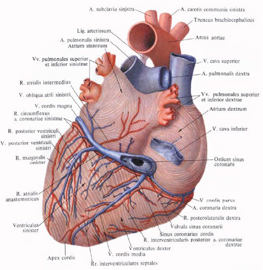
Here it sends twigs to the back wall of the right atrium and right ventricle (intermediate atrial branch, r. Atrialis intermedius), as well as thin branches, blood supplying the atrioventricular node and accompanying the atrioventricular bundle, branches of the atrioventricular node. Rr. Nodi atrioventricularis. On the diaphragmatic surface, it reaches the posterior interventricular furrow of the heart, in which it descends in the form of a posterior interventricular branch. R. Interventricularis posterior. The latter, approximately at the border of the middle and lower thirds of this furrow, plunges into the thickness of the myocardium. It supplies the posterior part of the interventricular septum (septal interventricular branches, interventricular septales) and the posterior walls of both the right and left ventricles.
At the place of transition of the main trunk into the interventricular furrow, a large branch leaves from it, passing along the coronary groove to the left half of the heart and feeding the posterior walls of the left atrium and left ventricle with its branches.
Left coronary artery, a. Coronaria sinistra, larger than the right one. Begins at the level of the left sinus of the aorta, follows left behind the root of the pulmonary trunk, and then between it and the left ear. Going to the left side of the coronal sulcus, even behind the pulmonary trunk is divided more often into two branches: the anterior interventricular branch and the envelope branch.

1. Anterior interventricular branch, r. Interventricularis anterior, is an extension of the main trunk. It descends the anterior interventricular sulcus to the apex of the heart, bends around it and enters the terminal section of the posterior interventricular sulcus; Not reaching the posterior interventricular branch, plunges into the thickness of the myocardium, giving off a number of septal interventricular branches, rr. Interventriculares septales. On the way, she sends twigs to the arterial cone (branch of the arterial cone, r. Soni arteriosi), to the nearby portions of the left and right ventricular walls, a larger branch to the anterior part of the interventricular septum, anastomotic branches to the trunks from the right coronary artery and completely blood supply to the apex Heart.
Near its beginning, the anterior interventricular branch gives a diagonally moving rather powerful lateral branch, r. Lateralis, which sometimes starts from the main trunk of the left coronary artery. In both cases, it branches out in the region of the anterior wall of the left ventricle.
2. The envelope branch, r. Circumflexus, leaving from under the left ear, follows the coronary groove to the pulmonary (lateral) surface of the heart and then along the posterior part of the coronal sulcus to the diaphragmatic surface of the heart, upon passage to which sends a large branch feeding the anterior and posterior walls of the left ventricle - the posterior Branch of left ventricle, r. Posterior ventriculi sinistri. Leaving from under the left ear, the artery gives off a large left marginal branch, r. Marginalis sinister, which follows down and somewhat posterior along the pulmonary (lateral) surface of the heart, heading toward the apex of the heart, and terminating in the anterior papillary muscle. Not reaching the posterior interventricular furrow, the envelope branch descends the diaphragmatic surface of the left ventricle, but does not reach the apex of the heart. On her way she sends twigs to the walls of the left ear and left atrium, which move away from the intermediate atrial branch, r. Atrialis intermedius, passing under a large vein of the heart to the diaphragm (bottom) surface of the left atrium. In addition, from the left coronary artery at the point of retreat of the posterior branch of the left ventricle the anastomatic atrial branch departs, r. Atrialis anastomoticus, which anastomoses with the branches of the right coronary artery in the venous sinus region.
Sometimes the envelope branch sends the unstable branches of the sinus-atrial and atrial-ventricular nodes, rr. Nodi sinuatrialis et atrioventricularis, anastomosing with the same branches from the right coronary artery.
Thus, the right coronary artery supplies the walls of the pulmonary trunk, the aorta, the right and left atria, the right ventricle, the posterior wall of the left ventricle, the interatrial and interventricular septa.
The left coronary artery supplies the walls of the pulmonary trunk, aorta, right and left auricles, the front walls of the right and left ventricles, the posterior wall of the left ventricle, the interatrial and interventricular septa.
The venous arteries of the heart are anastomosed with each other in all its parts, except for the right side and the pulmonary (lateral) surface of the heart, which are blood-sucked only by the corresponding arteries.
In addition, there are extra-anastomoses formed by vessels that feed the wall of the pulmonary trunk, the aorta and the hollow veins, as well as the vessels of the posterior wall of the atria. All these vessels anastomose with the arteries of the bronchi, the diaphragm and the pericardium.
In addition to intravenous anastomoses (intercoronary), anastomoses of branches of the same artery (intracoronary) are very well developed in the heart.
Intraorganic arteries of the heart, especially in the area of the ventricles, repeat the course of muscle beams: within the outer and deep layers of the myocardium, as well as papillary mucus arteries are directed along the longitudinal axis of the heart, and in the middle layer of the myocardium they have a transverse direction.

The veins .
Most of the veins of the heart, venae cordis (except for small and front), brings blood to a special reservoir of coronary sinus opening into the posterior part of the right atrial cavity, between the opening of the inferior vena cava and the right atrioventricular orifice.
The venous sinus, sinus coronarius, seems to be an extension to the diaphragmatic surface of the heart of his large vein. It is located in the left part of the posterior coronary furrow, from the place where it enters the upper oblique vein of the left atrium to its mouth: its length is 2-3 cm. A thin elephant of muscle beams of the myocardium is thrown over the coronary sinus, due to which its middle membrane , Tunica media.
The opening of the coronary sinus ostium sinus coronarii, in the cavity of the right atrium is bordered by a flap of the coronary sinus, valvula sinus coronarii. Two or three small flaps are located in the very sinus, near its opening.
The following veins belong to the coronary sinus system.
Great vein of the heart, v. Cordis magna, begins on the front surface of the apex of the heart. First, it lies in the anterior interventricular furrow next to the descending branch of the left coronary artery. Going up to the coronal sulcus, it is located in it and goes along the lower boundary of the left atrium to the pulmonary (lateral) surface of the heart. Rounding it, a large vein lies in the diaphragm part of the coronal sulcus, where it passes without a sharp boundary into the coronary sinus. Sometimes, at the site of the transition of the large vein of the heart into the coronary sinus, there is a small damper.
The veins of the anterior surface of both ventricles, the interventricular septum and sometimes near the sinus - the posterior vein of the left ventricle - enter the large vein of the heart.
1. The oblique vein of the left atrium, v. Obliqua atrii sinistri, begins on the lateral wall of the left atrium and goes from left to right downwards in the form of a small twig in the pericardial fold. Going down and to the right along the back wall of the left atrium, it passes into the coronary sinus. At the mouth of this vein, sometimes there is a small damper.
2. Posterior vein of left ventricle, v. Posterior ventriculi sinistri, originating on the posterolateral wall of the left ventricle, is directed upwards and falls into either a large vein of the heart or directly into the coronary sinus.
3. The average vein of the heart, v. Cordis media, begins on the diaphragm (bottom) surface in the region of the apex of the heart, passes in the posterior (lower) interventricular furrow next to the interventricular branch of the right coronary artery and flows into the right end of the coronary sinus. On the way it takes twigs from the diaphragm surface of both ventricles. In the area of the heart cutting, anastomosing with a large vein of the heart.
Small vein of the heart, v. Cordis parva, begins on the right side of the right atrium and right ventricle, passes in the posterior part of the coronary sulcus and flows either to the right end of the coronary sinus, or independently opens into the cavity of the right auricle, sometimes into the middle vein of the heart.
Outside the coronary sinus system, the following veins are described:
1. Fore veins of the heart, vv. Cordis anteriores, have a different value. They originate in the region of the anterior and lateral walls of the right ventricle, go upward and to the right to the coronary sulcus and flow directly into the right atrium; In the mouths of the anterior veins, there are sometimes small valves.
2. Least veins of the heart, vv. Cordis minimae, is a group of small veins collecting blood from various areas of the heart and opening with the opening of the smallest veins, foramina venarum minimarum, directly into the right and partly to the left atrium, as well as into the ventricles.

Comments
Commenting on, remember that the content and tone of your message can hurt the feelings of real people, show respect and tolerance to your interlocutors even if you do not share their opinion, your behavior in the conditions of freedom of expression and anonymity provided by the Internet, changes Not only virtual, but also the real world. All comments are hidden from the index, spam is controlled.