


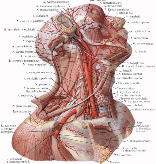
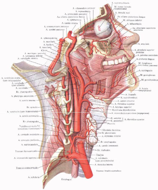
Arteries of the neck and head. External carotid artery
External carotid artery , a. Carotis externa, going up, goes somewhat ahead and medial to the internal carotid artery, and then outwards from it.
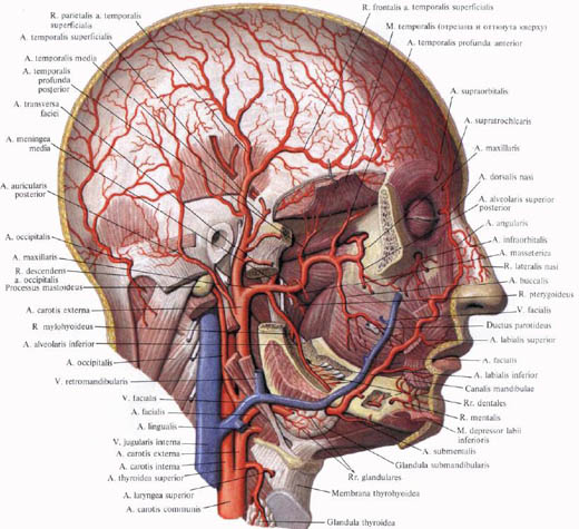
First, the external carotid artery is superficially covered by the subcutaneous muscle of the neck and the surface plate of the cervical fascia. Then, going up, passes behind the back abdomen of the digastric muscle and the sylvatic tubercle muscle. Somewhat higher it lies behind the branch of the lower jaw, where it penetrates into the parotid gland and at the level of the neck of the condylar process of the lower jaw is divided into the maxillary artery, a. Maxillaris, and superficial temporal artery, a. Temporalis superficialis, which form a group of terminal branches of the external carotid artery.
The external carotid artery gives a number of branches, which are divided into four groups: anterior, posterior, medial and a group of terminal branches.
Anterior group of branches. 1. Upper thyroid artery, a. Thyroidea superior, departs from the external carotid artery immediately at the site of the latter from the common carotid artery at the level of the large horn of the hyoid bone. It is directed slightly upward, then curved medially and follows the upper edge of the corresponding lobe of the thyroid gland, sending anterior glandular branch into its parenchyma, r. Glandularis anterior, posterior glandular branch, r. Glandularis posterior, and lateral glandular branch, r. Glandularis lateralis. In the thickness of the gland, the branches of the upper thyroid artery are anastomosed with the branches of the lower thyroid artery, a. Thyroidea inferior (from the thyroid shaft, truncus thyrocervicalis, escaping from the subclavian artery, a.subclavia).

In the course of the upper thyroid artery gives off a number of branches:
A) sub-lingual branch, r. Infrahyoideus, blood supply to the hyoid bone, and muscles attached to it; Anastomoses with the same branch of the opposite side;
B) sternocleidomastoid branch, r. Sternocleidomastoideus, impermanent, blood supply to the same muscle, approaching it from the inner surface, in its upper third;
C) upper laryngeal artery, a. Laryngea superior, goes to the medial side, passes over the upper edge of the thyroid cartilage, under the thyroid-lingual muscle and, perforating the thyroid-membrane, blood supply to the muscles, mucous membrane of the larynx and partly the hyoid bone and epiglottis:
D) cuspate branch, r. Cricothyroideus, blood supply to the same muscle and forms an arcuate anastomosis with an artery of the opposite side.

2. The lingual artery, a. Lingualis, thicker than the upper thyroid and starts slightly higher than it, from the anterior wall of the external carotid artery. In rare cases, it leaves the common trunk with the facial artery and is called the facial trunk, truncus linguofacialis. The lingual artery follows a little upward, passes over the large horn of the hyoid bone, heading forward and inside. In its turn, it is covered first by the abdominal back of the digastric muscle, the dorsal sublingual muscle, then passes under the sublingual-lingual muscle (between the last and middle constrictor of the pharynx from the inside), approaches the lower surface of the tongue , penetrating the thickness of its muscles.

In its turn, the lingual artery gives off a number of branches:
A) the sub-lingual branch, r. Suprahyoideus, passes along the upper edge of the hyoid bone, arched anastomoses with the same branch of the opposite side: blood supply to the hyoid bone and adjacent soft tissues;
B) dorsal branches of the tongue, rr. Dorsales linguae, of small thickness, move away from the lingual artery under the sublingual-lingual muscle, heading steeply upward, approaching the back of the tongue, blood supplying its mucous membrane and tonsil. Their terminal branches pass to the epiglottis and anastomose with the same arteries of the opposite side;
C) hyoid artery, a. Sublingualis, departs from the lingual artery until it enters the stratum of the tongue, is directed anteriorly, passing over the maxillofacial muscle outside of the mandibular duct; Further it approaches the hyoid gland, blood supplying it and a number of lying muscles; Ends in the mucosa of the bottom of the oral cavity and in the gum. A few twigs, perforating the jaw-hyoid muscle, anastomose with the sub-chanteral artery, a. Submentalis (branch of the facial artery, a. Facialis);
D) deep artery of the tongue, a. Profunda linguae, is the most powerful branch of the lingual artery, which is its extension. Going upwards, it enters into the thickness of the tongue between the chin-lingual muscle and the lower longitudinal muscle of the tongue; Then, following a twisty forward, reaches its top.
In its turn, the artery gives off numerous branches that feed their own muscles and the mucous membrane of the tongue. End branches of this artery approach the bridle of the tongue.
3. Facial artery, a. Facialis, originates from the anterior surface of the external carotid artery, somewhat higher than the lingual artery, is directed forward and upward and extends to the inside from the posterior abdomen of the digastric muscle and the sylvosvoid muscle into the submandibular jaw. Here it either adjoins the submaxillary gland, or perforates its thickness, and then it goes outward, skirting the lower edge of the body of the lower jaw in front of the attachment of the masticatory muscle; Bending upwards on the side surface of the face, approaches the area of the medial angle of the eye between the superficial and deep facial muscles.
In its turn, the facial artery gives off several branches:
A) ascending palatine artery, a. Palatina ascendens, departs from the initial section of the facial artery and, rising up the side wall of the pharynx, passes between the typhoid and shillopharyngeal muscles, supplying them with blood. The terminal branches of this artery branch into the pharyngeal opening of the auditory tube, in the tonsils and partially in the mucous membrane of the pharynx, where they anastomose with the ascending pharyngeal artery, a. Pharyngea ascendens;
B) amygdala branch, r. Tonsillaris, goes up the lateral surface of the pharynx, perforates the upper constrictor of the pharynx and ends with numerous branches in the thickness of the palatine tonsil. He gives a number of branches to the pharyngeal wall and the root of the tongue;
C) branches to the submaxillary gland - glandular branches, rr. Glandulares, are represented by several branches branching off from the main trunk of the facial artery in the place where it adjoins the submaxillary gland;
D) the sub-chordial artery, a. Submentalis, is a fairly powerful branch. Heading anteriorly, it passes between the anterior abdomen of the digastric muscle and the maxillo-hyoid muscle and circulates them. Anastomosing with the sublingual artery, the sub-chin artery passes through the lower crane of the lower jaw and, following the front face, blood supply to the skin and muscles of the chin and lower lip;
E) lower and upper labial arteries, aa. Labiales inferior and superior, start differently: the first - a little below the corner of the mouth, and the second - at the level of the angle, follow the thickness of the circular muscle of the mouth near the lip margin. The arteries supply blood to the skin, muscles and mucous membrane of the lips, anastomosing with the vessels of the opposite side with the same name. The upper labial artery gives off a thin branch of the septum of the nose, r. Septi nasi, blood supply to the skin of the septum of the nose in the region of the nostrils;
F) lateral branch of the nose, r. Lateralis nasi, - a small artery, is directed to the wing of the nose and blood supply to the skin of this area;
G) angular artery, a. Angularis, is the terminal branch of the facial artery. It goes up the side of the nose, giving small twigs to the wing and back of the nose. Then comes to the corner of the eye, where it is anastomosed with the dorsal artery of the nose, as well. Dorsalis nasi (branch of the ocular artery, a. Ophthlmica).
Rear group of branches. 1. The thoracic-clavicular-mastoid branch, r. Sternocleidomastoideus, often departs from the occipital artery or from the external carotid artery at the level of the beginning of the facial artery or somewhat higher and enters the thickness of the sternocleidomastoid muscle at the border of its middle and upper thirds.
2. Occipital artery, a. Occipitalis, is directed backwards and upwards. Initially, it is covered with the posterior abdomen of the digastric muscle and crosses the outer wall of the internal carotid artery. Then, under the posterior abdomen of the digastric muscle, it deviates posteriorly and proceeds in the furrow of the occipital artery of the mastoid process. Here, the occipital artery between the deep muscles of the occiput again goes upward and leaves medial to the place of attachment of the sternocleidomastoid muscle. Further, when the trapezius muscle is attached to the upper nasal line, it exits under the tendon helmet, where it gives off the terminal branches.
The following branches extend from the occipital artery:
A) sternocleidomastoid branches, rr. Sternocleidomastoidei, 3 to 4 blood supply the same muscle, as well as the surrounding muscles of the occiput; Sometimes move as a common trunk as a descending branch, r. Descendens;
B) mastoid branch, r. Mastoideus, - a thin trunk penetrating through the mastoid aperture to the dura mater;
C) ear, r. Auricularis, is directed forward and upward, blood supplying the back surface of the auricle;
D) occipital branches, rr. Occipitales, are terminal branches. Located between the supracranial muscle and the skin, they anastomose with each other and with the same branches of the opposite side, as well as with the branches of the posterior ear artery, as well. Auricularis posterior, and superficial temporal artery, a. Temporalis superficialis;
E) meningeal branch, r. Meningeus, - a thin trunk, penetrates through the parietal opening to the hard shell of the brain.
3. Posterior ear artery, a. Auricularis posterior, is a small vessel originating from the external carotid artery, above the occipital artery, but sometimes leaving it with a common trunk.
The posterior ear artery is directed upward, slightly posterior and inward and at first covered with the parotid gland. Then, ascending along the styloid process, it goes to the mastoid process, lying between it and the auricle. Here the artery divides into the anterior and posterior terminal branches.
A number of branches leave the posterior ear artery:
A) the stylomastor artery, a. Stylomastoidea, thin, passes through the same hole in the facial canal. Before entering the canal, a small artery - the posterior drum artery - departs from it, a. Tympanica posterior, penetrating into the tympanum through the stony-tympanic gap. In the canal of the facial nerve, it gives off small mastoid branches, rr. Mastoidei, to the cells of the mastoid process, and the stremal branch, r. Stapedialis, to the stremal muscle;
B) ear canal, r. Auricularis, passes over the back surface of the auricle and perforates it, giving twigs to the front surface;
C) occipital branch, r. Occipitalis, is guided along the base of the mastoid process back and forth, anastomosing with the terminal branches, as well. Occipitalis.
Medial group of branches.
Ascending pharyngeal artery, a. Pharyngea ascendens, begins from the inner wall of the external carotid artery. It is directed upward, goes between the inner and outer carotid arteries, approaches the side wall of the pharynx.
Returns the following branches:
A) pharyngeal branches, rr. Pharyngeales, two or three, are guided along the back wall of the pharynx and supply the back of the pharynx with the palatine tonsil to the base of the skull, as well as part of the soft palate and partly the auditory tube;
B) posterior meningeal artery, a. Meningea posterior, follows up the internal carotid artery, as well. Carotis interna, or through the jugular opening; Further passes into the cavity of the skull and branches into the hard shell of the brain;
C) lower drum artery, a. Tympanica inferior, - a thin stem that penetrates into the tympanum through the tympanic tubule and blood supply to its mucous membrane.
Group of terminal branches.
I. Maxillary artery, a. Maxillaris, departs from the external carotid at a right angle at the level of the neck of the lower jaw. The initial section of the artery is covered with the parotid gland. Then the artery, wriggling, is directed horizontally anteriorly between the branch of the lower jaw and the wedge-mandibular ligament.
Further, the artery passes between the lateral pterygoid muscle and the temporal muscle and reaches the pterygopalatine fossa, where it divides into terminal branches.
Branches that depart from the maxillary artery, respectively, the topography of its individual sites are conventionally divided into three groups.
The first group includes branches that depart from the main trunk a. Maxillaris near the neck of the lower jaw - these are the branches of the mandibular part of the maxillary artery.
The second group consists of branches starting from that department a. Maxillaris, which lies between the lateral pterygoid and temporal muscles, are the branches of the pterygoid part of the maxillary artery.
The third group includes branches that depart from that section of a. Maxillaris, which is located in the pterygoid-palatine fossa, is the branch of the pterygoid-palatal part of the maxillary artery.
Branch of the lower jaw part. 1. Deep ear canal, a. Auricularis profunda, is a small branch that extends from the initial section of the main trunk. It is directed upward and blood supply to the joint capsule of the temporomandibular joint, the lower wall of the external auditory canal and the tympanic membrane.
2. Anterior drum artery, a. Tympanica anterior, often a branch of the deep ear artery. Penetrates through the stony-tympanic gap into the tympanum cavity, blood supplying its mucous membrane.
3. Lower alveolar artery, a. Alveolaris inferior, a fairly large vessel, is directed downward, entering through the opening of the lower jaw into the canal of the lower jaw, where it passes along with the same vein and nerve. In the canal from the artery the following branches branch out:
A) dental branches, rr. Dentales, passing into more subtle teeth;
B) toothed branches, rr. Peridentales, suitable for teeth, periodontium, dental alveoli, gum, spongy substance of the lower jaw;
C) maxillo-hyoid branch, r. Mylohyoideus, departs from the lower alveolar artery before its entry into the canal of the lower jaw, goes in the maxillofacial groove and blood supply to the maxillofacial muscle and the anterior abdomen of the digastric muscle;
D) chin branch, r. Mentalis, is an extension of the lower alveolar artery. Exits through the chin opening on the face, disintegrating into a number of branches, blood supply to the area of the chin and lower lip and anastomoses with branches a. Labialis inferior and a. Submentalis.
Branch of the pterygoid part.
1. The middle meningeal artery, a. Meningea media, is the largest branch that extends from the maxillary artery. It is directed upward, passes through the spinous aperture into the cranial cavity, where it divides into the frontal and parietal branches, rr. Frontalis et parietalis. The latter go along the outer surface of the hard shell of the brain in the arterial grooves of the bones of the skull, supplying them with blood, and also the temporal, frontal and parietal parts of the membrane.
In the course of the middle meningeal artery, the following branches depart from it:
A) the upper drum artery, a. Tympanica superior, - a thin vessel; Entering through the cleft of the channel of the small stony nerve into the tympanum cavity, blood supply to its mucous membrane;
B) stony branch, r. Petrosus, originates above the awned hole, follows laterally and posteriorly, enters the cleft of the channel of the large stony nerve. Here it is anastomosed with the branch of the posterior ear artery - the stylomastor artery, a. Stylomastoidea;
C) orbital branch, r. Orbitalis, thin, is directed anteriorly and, accompanying the optic nerve, enters the orbit;
D) an anastomotic branch (with a lachrymal artery), r. Anastomoticus (cum a. Lacrimali), penetrates through the upper orbital fissure into the orbit and anastomoses with the lachrymal artery, a. Lacrimalis, - branch of the eye artery;
E) pterygoid-meningeal artery, a. Pterygomeningea, departs still outside the cranial cavity, blood supply to the pterygoid muscles, auditory tube, muscles of the sky. Having entered through the oval aperture into the cavity of the skull, blood supply to the trigeminal node. Can depart directly from a. Maxillaris, if the latter lies not on the lateral, but on the medial surface of the lateral pterygoid muscle.
2. Deep temporal arteries, aa. Temporales profundae, represented by the anterior deep temporal artery, as well. Temporalis profunda anterior, and posterior deep temporal artery, a. Temporalis profunda posterior. They move away from the main trunk of the maxillary artery, they go upward into the temporal fossa, lying between the skull and the temporal muscle, and supply the deep and lower parts of this muscle.
3. Chewing artery, a. Masseterica, sometimes originating from the posterior deep temporal artery and passing through the incision of the lower jaw onto the outer surface of the lower jaw, approaches the chewing muscle from the side of its inner surface, blood supplying it.
4. Posterior alveolar artery, a. Alveolaris superior posterior, begins near the hillock of the upper jaw with one or two - three branches. Going down, penetrates through the alveolar foramina in the epithelium of the same name, where it gives off the dental branches, rr. Dentales, passing into the peri-toothed branches, rr. Peridentales, reaching the roots of large molars of the upper jaw and gums.
5. Genital artery, a. Buccalis, - a small vessel, directed forward and downward, passes through the buccal muscle, blood supply to it, the mucous membrane of the mouth, gums in the upper teeth and a number of nearby mimic muscles. Anastomoses with the facial artery.
6. Pterygoid branches, rr. Pterygoidei, only 2 - 3, are directed to the lateral and medial pterygoid muscles.
Branches of the pterygoid-palatine part. 1. The infraorbital artery, a. Infraorbitalis, passes through the lower orbital gap into the orbit and goes in the infraorbital furrow, then passes through the eponymous canal and through the infraorbital foramina emerges to the surface of the face, giving the terminal branches to the tissues of the infraorbital area of the face.
On its way, the infraorbital artery sends the anterior superior alveolar arteries, aa. Alveolares superiores anteriores, which pass through the canals in the outer wall of the maxillary sinus and, connecting with the branches of the posterior superior alveolar artery, render the dental branches, rr. Dentales, and peri-toothed branches, rr. Peridentales, directly blood supplying the teeth of the upper jaw, gum and mucous membrane of the maxillary sinus.
2. Descending palatine artery, a. Palatina descendens, in its initial department gives the artery of the pterygoid canal, a. Canalis pterygoidei (can depart independently, giving the pharyngeal branch, r. Pharyngeus), it goes downwards, it penetrates into the large palatine canal and divides into small and large palatine arteries, aa. Palatinae minores et major, and unstable pharyngeal branch, r. Pharyngeus. Small palatine arteries pass through a small palatine hole and supply blood to the soft palate and palatine tonsil. A large palatine artery emerges from the canal through a large palatine foramen, goes into the palatal fissure of the hard palate; Blood supply to its mucous membrane, glands and gum; Going forward, passes up through the incisive canal and anastomoses with the posterior septum branch, r. Septalis posterior. Some branches anastomose with the ascending palatine artery, a. Palatina ascendens, - branch of the facial artery, a. Facialis.
3. Wedgly-palatine artery, a. Sphenopalatina, - terminal vessel of the maxillary artery. Passes through the wedge-palatine foramen in the nasal cavity and divides here into a series of branches:
A) lateral posterior nasal arteries, aa. Nasales posteriores laterales, - fairly large branches, cover the mucous membrane of the middle and lower shells, the lateral wall of the nasal cavity and terminate in the mucous membrane of the frontal and maxillary sinuses;
B) rear partitions, rr. Septales posteriors, are divided into two branches (upper and lower), blood supply to the mucosa of the septum of the nose. These arteries, moving forward, anastomose with the branches of the eye artery (from the internal carotid), and in the incisors area - with a large palatine artery and artery of the upper lip.
II. Superficial temporal artery, a. Temporalis superficialis, - the second terminal branch of the external carotid artery, which is its continuation. It originates in the neck of the lower jaw.
It is directed upwards, passes in the thickness of the parotid gland between the external ear canal and the head of the lower jaw, then, lying superficially under the skin, follows the root of the zygomatic arch, where it can be probed. Somewhat higher than the zygomatic artery, the artery divides into its terminal branches: the frontal branch, r. Frontalis, and parietal branch, r. Parietalis.
In its turn, the artery gives off a series of branches.
1. The branches of the parotid gland, rr. Parotidei, only 2 - 3, blood supply to the parotid gland.
2. The transverse artery of the face, a. Transversa facialis, is initially located in the thickness of the parotid gland, blood supplying it, then passes horizontally along the surface of the masticatory muscle between the lower edge of the zygomatic arch and the parotid duct, giving twigs to mimic muscles and anastomosing with branches of the facial artery.
3. Front ear branches, rr. Auriculares anteriores, only 2-3, are directed to the anterior surface of the auricle, blood supplying its skin, cartilage and muscle.
4. The middle temporal artery, a. Temporalis media, going upwards, perforates the temporal fascia above the zygomatic arch (from the surface into the depth) and enters the thickness of the temporal muscle and blood supply it.
5. Scutiocular artery, a. Zygomaticoorbitalis, is directed above the zygomatic arch forward and upward, reaching the circular muscle of the eye. It supplies a number of facial muscles and anastomoses with a. Transversa facialis, r. Frontalis and a. Lacrimalis of a. Ophthalmica.
6. Frontal branch, r. Frontalis, one of the terminal branches of the superficial temporal artery, is directed forward and upward and blood supply to the frontal abdomen of the occipitus-frontal muscle, the circular muscle of the eye, the tendon helmet and the forehead skin.
7. The dark branch, r. Parietalis, - the second terminal branch of the superficial temporal artery, somewhat larger than the frontal branch. It goes up and back, blood supply to the skin of the temporal region; Anastomosis with the same branch of the opposite side.

Comments
When commenting on, remember that the content and tone of your message can hurt the feelings of real people, show respect and tolerance to your interlocutors even if you do not share their opinion, your behavior in the conditions of freedom of expression and anonymity provided by the Internet, changes Not only virtual, but also the real world. All comments are hidden from the index, spam is controlled.