


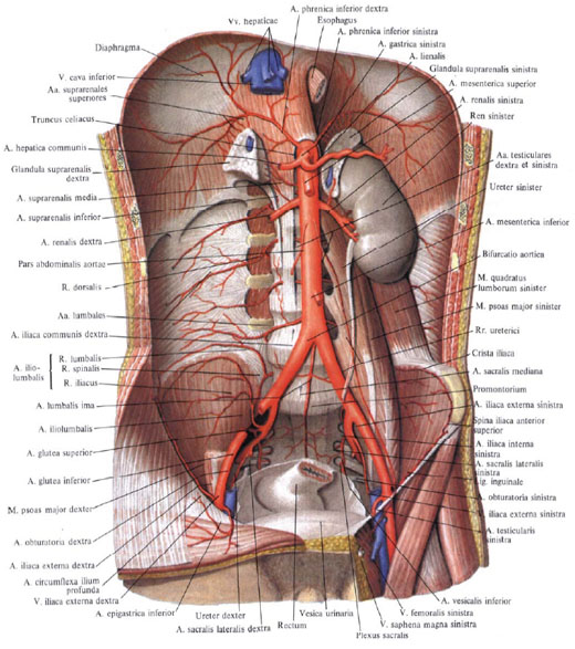
Abdominal part of the aorta
The abdominal part of the aorta (abdominal aorta), pars abdominalis aortae (aorta abdominalis), is the continuation of the thoracic part of the aorta. It begins at the level of the XII thoracic vertebra and reaches the IV-V of the lumbar vertebra. Here the abdominal aorta is divided into two common iliac arteries, aa. Aliacae communes. The place of division is called bifurcation of the aorta, bifurcatio aortica. From bifurcation downward a thin twig lying on the anterior surface of the sacrum, - the median sacral artery, a. Sacralis mediana.
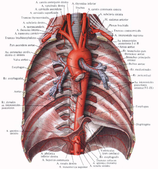
From the ventral part of the aorta there are two kinds of branches: parietal and internal.
The abdominal part of the aorta is located retroperitoneally. In the upper part of its surface, adjoining, crossing it, the body of the pancreas and two veins: the splenic vein lying along the upper edge of the pancreas, v. Lienalis, and left renal vein, v. Renalis sinistra, going behind the gland. Below the body of the pancreas, in front of the aorta, is the lower part of the duodenum, and below it - the beginning of the root of the mesentery of the small intestine. To the right of the aorta lies the inferior vena cava, v. Cava inferior; Behind the initial abdominal aorta is the cistern of the thoracic duct, cisterna chyli, the initial part of the thoracic duct, ductus thoracicus.
Parietal branches.
1. Lower diaphragmatic artery, a. Phrenica inferior, - quite powerful pair artery. It departs from the anterior surface of the initial part of the abdominal aorta at the level of the XII thoracic vertebra and is directed to the lower surface of the tendon part of the diaphragm where it gives the front and back branches that supply the latter. In the thickness of the diaphragm, the right and left arteries are anastomosed with each other and with branches from the thoracic part of the aorta. The right artery passes behind the inferior vena cava, the left artery behind the esophagus.
In its turn, the artery gives 5-7 upper adrenal arteries, aa. Suprarenales superiores. These are thin branches that depart from the initial section of the lower diaphragmatic artery and supply blood to the adrenal gland. On the way from them several small branches run to the lower parts of the esophagus and to the peritoneum.
2. Lumbar arteries, aa. Lumbales, represent 4 paired arteries. They move away from the posterior wall of the abdominal part of the aorta at the level of the body I-IV of the lumbar vertebrae. They are directed transversely, laterally, while the two upper arteries pass behind the diaphragm legs, the two lower - behind the large lumbar muscle.
All lumbar arteries anastomose with each other and with the upper and lower epigastric arteries, blood supplying the rectus abdominis muscle. In its course, the arteries give a series of small branches to the subcutaneous tissue and to the skin; In the field of the white line they anastomose in some places with the same arteries of the opposite side. In addition, lumbar arteries are anastomosed with intercostal arteries, aa. Intercostales, ilio-lumbar artery, a. Iliolumbalis, the deep artery surrounding the iliac bone, a. Circumflexa ilium profunda, and the upper gluteal artery, a. Glutea superior.
Having reached the transverse processes of the vertebrae, each lumbar artery gives off the dorsal branch, r. Dorsalis. Then the lumbar artery goes behind the square muscle of the waist, blood supplying it; Then goes to the front wall of the abdomen, passes between the transverse and internal oblique muscles of the abdomen and reaches the rectus abdominis muscle.
The dorsal branch goes to the back surface of the trunk to the back muscles and the skin of the waist region. On the way, she gives a small branch to the spinal cord - the spinal branch, r. Spinalis, which enters through the intervertebral opening into the vertebral canal, blood supplying the spinal cord and its membranes.

3. The median sacral artery, a. Sacralis mediana, is a direct extension of the abdominal aorta. It starts from the posterior surface, slightly above the aortic bifurcation, ie, at the level of the V lumbar vertebra. It is a thin vessel that runs from the top down in the middle of the pelvic surface of the sacrum and ends on the coccyx in the coccygeal glomus coccygeum.
From the median sacral artery along the course of it branch off:
A) inferior lumbar artery, a. Lumbalis imae, steam, departs in the region of the V lumbar vertebra and blood supply to the ilio-lumbar muscle. On its way, the artery gives the dorsal branch involved in the blood supply to the deep muscles of the back and spinal cord;
B) lateral sacral branches, rr. Sacrales laterales, move away from the main trunk at the level of each vertebra and, branching on the anterior surface of the sacrum, anastomose with similar branches from the lateral sacral arteries (branches of internal iliac arteries).
From the lower part of the median sacral artery leaves a few branches that supply blood to the lower parts of the rectum and loose fiber around it.
Internal branches
I. The trunk , truncus celiacus, is a short vessel 1 to 2 cm long, extending from the anterior surface of the aorta at the level of the upper edge of the body I of the lumbar vertebra or the lower edge of the body of the XII thoracic vertebra where the abdominal aorta emerges from the aortic orifice. The artery goes anteriorly and is immediately divided into three branches: the left gastric artery, a. Gastricasinistra, a common hepatic artery, a. Hepatica communis, and splenic artery, a. Splenica (lienalis).

1. Left gastric artery, a. Gastrica sinistra, the smaller of the three arteries. It rises a little up and to the left; Going to the cardiac part of the stomach , gives a few branches towards the esophagus - esophageal branches, rr. Esophageales, anastomosing with the same branches from the thoracic part of the aorta, and itself descends to the right side by a small curvature of the stomach, anastomosing with the right gastric artery, a. Gastrica dextra (from the common hepatic artery). On its way along the small curvature, the left gastric artery sends small twigs to the anterior and posterior walls of the stomach.
2. The common hepatic artery, a. Hepatica communis, a more powerful branch, has a length of up to 4 cm. Moving away from the celiac trunk, it follows the right foot of the diaphragm, the upper edge of the pancreas from left to right and enters the thickness of the small omentum, where it divides into two branches - its own hepatic and gastroduodenal arteries.
1) Own hepatic artery, a. Hepatica propria, moving away from the main trunk, goes to the gates of the liver in the thickness of the hepatic-duodenal ligament, to the left of the common bile duct and somewhat anterior to the portal vein, v. Portae. Going to the gates of the liver, your own hepatic artery divides into the left and right branches, with the right jaw branching the cholelithiasis artery, a. Cystica.
The right gastric artery, a. Gastrica dextra, - a thin branch, departs from its own hepatic artery, sometimes from the common hepatic artery. It is directed from the top down to the small curvature of the stomach, along which it goes from right to left, and anastomoses with a. Gastrica sinistra. The right gastric artery gives a number of branches, blood supplying the anterior and posterior walls of the stomach.
In the gates of the liver, the right branch, r. Dexter, its own hepatic artery sends to the caudate lobe the artery of the caudate lobe, a. Lobi caudati, and arteries to the corresponding segments of the right lobe of the liver: to the anterior segment - the artery of the anterior segment, a. Segmenti anterioris, and to the posterior segment - the artery of the posterior segment, a. Segmenti posterioris.
Left branch, r. Sinister, gives the following arteries: the artery of the caudate lobe, a. Lobi caudati, and the arteries of the medial and lateral segments of the left lobe of the liver, a. Segmenti medialis et a. Segmenti lateralis. In addition, from the left branch (less often from the right branch) a non-stationary intermediate branch departs, r. Intermedius, blood supplying the square share of the liver.
2) The gastroduodenal artery, a. Gastroduodenalis, is a fairly powerful trunk. It is directed from the common hepatic artery downwards, behind the pylorus part of the stomach, crossing it from the top down. Sometimes a superduodenal artery departs from this artery, a. Supraduodenalis, which crosses the anterior surface of the head of the pancreas.
The following branches leave the gastroduodenal artery:
A) posterior upper pancreatoduodenal artery, a. Pancreaticoduodenalis superior posterior, runs along the posterior surface of the head of the pancreas, and, going down, gives on its move pancreatic branches, rr. Pancreatici, and duodenal branches, rr. Duodenales. At the lower edge of the horizontal part of the duodenum, the artery is anastomosed with the lower pancreatoduodenal artery, a. Pancreaticoduodenalis inferior (branch of the superior mesenteric artery, a. Mesenterica superior);
B) anterior superior pancreatoduodenal artery, a. Pancreaticoduodenalis superior anterior, located arched on the front surface of the head of the pancreas and the medial edge of the descending portion of the duodenum, directed downward, giving up in its path the duodenal branches, rr. Duodenales, and pancreatic branches, rr. Pancreatici. At the lower edge of the horizontal part of the duodenum anastomoses with the lower pancreatoduodenal artery, a. Pancreatoduodenalis inferior (branch of the superior mesenteric artery).
C) right gastro-omental artery, a. Gastroepiploica dextra, is an extension of the gastroduodenal artery. Sent to the left along the large curvature of the stomach between the leaves of the large omentum, sends twigs to the anterior and posterior walls of the stomach - the gastric branches, rr. Gastrici, as well as glandular branches, rr. Epiploici to the large stuffing box. In the region of great curvature, it is anastomosed with the left gastro-omental artery, a. Gastroepiploica sinistra (branch of the splenic artery, a. Splenica);
D) pre-duodenal arteries, aa. Retroduodenales, are the right terminal branches of the gastroduodenal artery. They surround the front surface of the right edge of the head of the pancreas.

3. Splenic artery, a. Splenica, is the thickest of the branches that extend from the celiac trunk. The artery goes to the left and, together with the eponymous vein, lies behind the upper edge of the pancreas. Reaching the tail of the pancreas, enters the gastro-splenic ligament and splits into terminal branches that are directed towards the spleen.
Splenic artery gives branches, blood supply to the pancreas, stomach and a large omentum.
1) Pancreatic branches, rr. Pancreatici, depart from the splenic artery throughout its entire length and enter the parenchyma of the gland. They are represented by the following arteries:
A) dorsal pancreatic artery, a. Pancreatica dorsalis, follows the middle section of the posterior surface of the body of the pancreas and at the lower edge of the pancreas passes into the lower pancreatic artery, a. Pancreatica inferior, blood supplying the lower surface of the pancreas;
B) a large pancreatic artery, a. Pancreatica magna, departs from the main trunk or from the dorsal pancreatic artery, it follows to the right and goes along the back surface of the body and head of the pancreas. It connects with the anastomosis between the posterior upper and lower pancreatoduodenal arteries;
C) caudal pancreatic artery, a. Caude pancreatis, is one of the terminal branches of the splenic artery, blood supply to the tail of the pancreas.
2) Splenic branches, rr. Splenici, only 4 to 6, are the terminal branches of the splenic artery and penetrate through the gates into the parenchyma of the spleen.
3) Short gastric arteries, aa. Gastricae breves, in the form of 3 -7 small stems depart from the terminal segment of the splenic artery and in the thickness of the gastro-splenic ligament go to the bottom of the stomach, anastomosing with other gastric arteries.
4) Left gastro-omental artery, a. Gastroepiploica sinistra, begins from the splenic artery in the place where the terminal branches from the spleen leave from it, and it follows down in front of the pancreas. Having reached the big curvature of the stomach, it is guided along it from left to right, lying between the leaves of the large omentum. At the border of the left and middle thirds of the large curvature anastomoses with the right gastro-omental artery (from the gastroduodenalis). In its turn, the artery sends a number of branches to the anterior and posterior walls of the stomach - gastric vetvi, rr. Gastrici, and to the large epiploon - stuffing branches, rr. Epiploici.
5) Posterior gastric artery, a. Gastrica posterior, impermanent, blood supply to the back wall of the stomach, closer to the cardial part.
II. Upper mesenteric artery , a. Mesenterica superior, is a large vessel that starts from the anterior surface of the aorta, slightly below (by 1 to 3 cm) the celiac trunk, behind the pancreas.

Leaving from beneath the lower edge of the gland, the superior mesenteric artery goes down and to the right. Together with the upper mesenteric vein located to the right of it, runs along the anterior surface of the horizontal (ascending) part of the duodenum, it crosses it right to the right of the duodenum-lean curve. Having reached the root of the mesentery of the small intestine, the superior mesenteric artery penetrates between the leaves of the latter, forming an arch facing the bulge to the left, and reaches the right ileal fossa.
In its turn, the superior mesenteric artery gives the following branches: to the small intestine (with the exception of the upper part of the duodenum), to the caecum with a vermiform appendix, ascending and partly to the transverse colon.
The following arteries extend from the superior mesenteric artery.
1. Lower pancreatoduodenal artery, a. Pancreaticoduodenalis inferior (sometimes unequal), originates from the right edge of the initial segment of the superior mesenteric artery. It is divided into an anterior branch, r. Anterior, and posterior branch, r. Posterior, which are sent down and to the right along the front surface of the pancreas, round its head along the border with the duodenum. Gives twigs to the pancreas and duodenum; Anastomosing with anterior and posterior upper pancreatoduodenal arteries and with branches a. Gastroduodenalis.
2. Articulate arteries, aa. Jejunales, 7 to 8 in all, depart one by one from the convex part of the arch of the superior mesenteric artery, between the leaves of the mesentery and the loins of the jejunum. On its way, each branch is divided into two trunks that anastomose with the same trunks formed from the division of adjacent intestinal arteries.
3. The ileo-intestinal arteries, aa. Ileales, in the number of 5 to 6, like the previous ones, are directed to the loops of the ileum and, dividing into two trunks, anastomose with a number of going intestinal arteries. Such anastomoses of the intestinal arteries look like arches. From these arcs new branches branch out, which also divide, forming arcs of the second order (somewhat smaller magnitude). From arcs of the second order arteries again leave, which, dividing, form arcs of the third order, etc. From the last, most distal series of arcs, straight branches directly go to the walls of the loops of the small intestine. In addition to intestinal loops, these arches give small twigs, blood supply mesenteric lymph nodes.
4. The iliac-colon intestinal artery, a. Ileocolica, departs from the cranial half of the superior mesenteric artery. Heading to the right and down under the parietal peritoneum of the back wall of the abdominal cavity to the end of the ileum and to the cecum, the artery divides into branches that supply the cecum, the beginning of the colon and the terminal part of the ileum.
From the ileo-colonic artery leaves a number of branches:
A) the ascending artery goes to the right to the ascending colon, rises along its medial edge and anastomizes (forms an arc) with the right colon-intestinal artery, a. Colica dextra. From the indicated arch, branch-intestinal branches leave, rr. Colici, blood supply to the ascending colon and upper part of the cecum;
B) anterior and posterior cecal arteries, aa. Cecales anterior and posterior, are directed to the corresponding surfaces of the caecum. They are a continuation of a. Ileocolica, approach the ileocecal angle, where, connecting with the terminal branches of the ileo-intestinal arteries, form an arc from which the branches branch to the caecum and to the terminal part of the ileum, iliac-intestinal branches, rr. Ileales;
C) arteries of the appendix, aa. Appendiculares, depart from the posterior cecal artery between the leaves of the mesentery of the appendix; Blood supply vveroobrazny process.
5. Right colon-intestinal artery. A. Colica dextra, departs from the right side of the superior mesenteric artery, in its upper third, at the root level of the mesentery of the transverse colon, and is directed almost transversely to the right, to the medial edge of the ascending colon. Not reaching the ascending colon, is divided into the ascending and descending branches. The descending branch joins the branch a. Ileocolica, and the ascending branch anastomoses with the right branch a. Colica media. From the arcs formed by these anastomoses, branch off to the wall of the ascending colon, to the right bend of the colon and to the transverse colonic intestine.
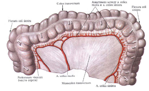
6. The average colon-intestinal artery, a. Colica media, departs from the initial section of the superior mesenteric artery, is directed forward and to the right between the leaves of the mesentery of the transverse colon and is divided at the bottom of the branch: the right and left.
The right branch connects with the ascending branch a. Colica dextra, and the left branch goes along the mesenteric margin of the transverse colon and anastomizes with the ascending branch of a. Colica sinistra, which departs from the inferior mesenteric artery. Joining this way with the branches of neighboring arteries, the middle colonic artery forms arcs. From the branches of these arcs arcs of the second and third order are formed, which give straight branches to the walls of the transverse colon, to the right and left flexures of the colon.
III. Lower mesenteric artery , a. mesenterica inferior, отходит от передней поверхности брюшной аорты на уровне нижнего края III поясничного позвонка. Артерия идет позадибрюшинно влево и вниз и разделяется на три ветви.

1. Левая ободочно-кишечная артерия, a. colica sinistra, залегает забрюшинно в левом брыжеечном синусе впереди левого мочеточника и левой яичковой (яичниковой) артерии, а. testicularis (ovarica) sinistra; разделяется на восходящую и нисходящую ветви. Восходящая ветвь анастомозирует с левой ветвью средней ободочно-кишечной артерии, образуя дугу; кровоснабжает левую часть поперечной ободочной кишки и левый изгиб ободочной кишки. Нисходящая ветвь соединяется с сигмовидно-кишечной артерией и кровоснабжает нисходящую ободочную кишку.
2. Сигмовидно-кишечная артерия, a. sigmoidea (иногда их несколько), идет вниз сначала забрюшинно, а затем между листками брыжейки сигмовидной ободочной кишки; анастомозирует с ветвями левой ободочно-кишечной артерии и верхней прямокишечной артерии, образуя дуги, от которых отходят ветви, кровоснабжающие сигмовидную ободочную кишку.
3. Верхняя прямокишечная артерия, а. rectalis superior, является концевой ветвью нижней брыжеечной артерии; направляясь вниз, разделяется на две ветви. Одна ветвь анастомозирует с ветвью сигмовиднокишечной артерии и кровоснабжает нижние отделы сигмовидной ободочной кишки. Другая ветвь направляется в полость малого таза, пересекает спереди a. iliaca communis sinistra и, залегая в брыжейке тазового отдела сигмовидной ободочной кишки, разделяется на правую и левую ветви, которые кровоснабжают ампулу прямой кишки. В стенке кишки они анастомозируют со средней прямокишечной артерией, а. rectalis media, ветвью внутренней подвздошной артерии, a. Iliaca interna.
IV. Средняя надпочечниковая артерия , a. suprarenalis media, парная, отходит от боковой стенки верхнего отдела аорты, несколько ниже места отхождения брыжеечной артерии. Направляется поперечно кнаружи, пересекает ножку диафрагмы и подходит к надпочечнику, в паренхиме которого анастомозирует с веточками верхней и нижней надпочечниковых артерий.
V. Почечная артерия , a. renalis,— парная крупная артерия. Начинается от боковой стенки аорты на уровне II поясничного позвонка почти под прямым углом к аорте, на 1—2 см ниже отхождения верхней брыжеечной артерии. Правая почечная артерия несколько длиннее левой, так как аорта лежит слева от срединной линии; направляясь к почке, она располагается позади нижней полой вены.
Не доходя до ворот почки, каждая почечная артерия отдает небольшую нижнюю надпочечниковую артерию, a. suprarenalis inferior, которая, проникнув в паренхиму надпочечника, анастомозирует с ветвями средней и верхней надпочечниковых артерий.
В области ворот почки почечная артерия делится на переднюю и заднюю ветви.
Передняя ветвь, r. anterior, входит в почечные ворота, проходя впереди почечной лоханки, и ветвится, посылая артерии к четырем сегментам почек: артерию верхнего сегмента, a. segmenti superioris, — к верхнему; артерию верхнего переднего сегмента, a. segmenti anterior superioris, — к верхнему переднему; артерию нижнего переднего сегмента, а. segmenti anterior is inferioris, — к нижнему переднему и артерию нижнего сегмента, a. segmenti inferioris, — к нижнему. Задняя ветвь, r. posterior, почечной артерии проходит позади почечной лоханки и, направляясь в задний сегмент, отдает мочеточниковую ветвь, r. uretericus, которая может отходить от самой почечной артерии, разделяется на заднюю и переднюю веточки.
VI. Яичковая артерия , a. testicularis, парная, тонкая, отходит (иногда правая и левая общим стволом) от передней поверхности брюшной аорты, несколько ниже почечной артерии. Направляется вниз и латерально, идет по большой поясничной мышце, пересекает на своем пути мочеточник, над дугообразной линией — наружную подвздошную артерию. По пути отдает веточки к жировой капсуле почки и к мочеточнику — мочеточниковые ветви, rr. ureterici. Далее направляется к глубокому паховому кольцу и, присоединившись здесь к семявыносящему протоку, переходит через паховый канал в мошонку и распадается на ряд мелких веточек, идущих в паренхиму яичка и его придатку, — ветви придатка яичка, rr. epididymales.
По своему ходу анастомозирует с a. cremasterica (ветвь a. epigastrica inferior и с a. ductus deferentis (ветвь а. iliaca interna).
У женщин соответствующая яичковой артерии яичниковая артерия, а. ovarica, отдает ряд мочеточниковых ветвей, rr. ureterici, а затем проходит между листками широкой связки матки, вдоль ее свободного края, и отдает ветви к маточной трубе — трубные ветви, rr. tubales, и в ворота яичника. Концевая ветвь яичниковой артерии анастомозирует с яичниковой ветвью маточной артерии.

Comments
When commenting on, remember that the content and tone of your message can hurt the feelings of real people, show respect and tolerance to your interlocutors even if you do not share their opinion, your behavior in the conditions of freedom of expression and anonymity provided by the Internet, changes Not only virtual, but also the real world. All comments are hidden from the index, spam is controlled.