Arteries of the lower extremity. Femoral artery
Femoral artery , a. Femoralis, is a continuation of the external iliac artery and begins under the inguinal ligament in the vascular lacunae. Femoral artery, coming out on the front surface of the thigh, is directed downward and medially, lying in the groove between the anterior and medial groups of the muscles of the thigh. In the upper third of the artery is located within the femoral triangle, on a deep leaf of the broad fascia, covered by its surface leaf; The femoral vein passes medially from it. Passing the femoral triangle, the femoral artery (along with the femoral vein) is covered by a sartorius muscle and enters the upper opening of the leading canal at the border of the middle and lower thirds of the thigh. In this canal, the artery is located along with the subcutaneous nerve, n. Saphenus, and femoral vein, v. Femoralis. Together with the latter, it deflects posteriorly and emerges through the lower opening of the canal to the posterior surface of the lower limb into the popliteal fossa, where it receives the name of the popliteal artery, a. Poplitea.
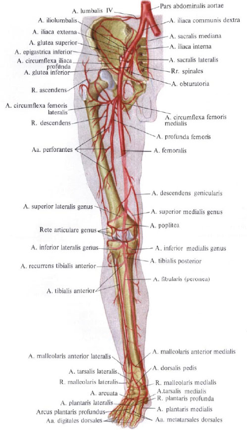
The femoral artery gives off a series of branches, blood supplying the thigh and the anterior wall of the abdomen.
1. Superficial epigastric artery, a. Epigastrica superficialis, begins from the anterior wall of the femoral artery below the inguinal ligament, perforates the superficial leaf of the broad fascia in the area of the subcutaneous fissure, and, rising upward and medially, passes to the anterior abdominal wall where it lies subcutaneously and reaches the region of the umbilical ring. Here its branches anastomose with branches a. Epigastrica superior (from a thoracica interna). The branches of the superficial epigastric artery supply the skin of the anterior abdominal wall and the external oblique muscle of the abdomen.
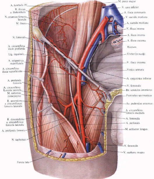
2. The superficial artery surrounding the iliac bone, a. Circumflexa iliaca superficialis, departs from the external wall of the femoral artery or from the superficial epigastric artery and is guided laterally along the inguinal ligament to the superior anterior iliac spine; Blood supply to the skin, muscles and inguinal lymph nodes.
3. External genitalia, aa. Pudendae externae, in the form of two, sometimes three thin stems are guided medially, skirting the anterior and posterior periphery of the femoral vein. One of these arteries goes up and reaches the suprapubic region, branched into the skin. Other arteries, passing over the crest muscle, perforate the fascia of the femur and approach the scrotum (labia) - these are the front scrotal (labial) branches, rr. Scrotales (labiales) anteriores.
4. Inguinal branches, rr. Inguinales, depart from the initial section of the femoral artery or from the external genital arteries (3-4) with small trunks and, perforating the wide fascia of the thigh in the region of the latticed fascia, blood supply the skin, as well as superficial and deep lymph nodes of the inguinal region.
5. Deep thigh artery, a. Profunda femoris, is the most powerful branch of the femoral artery. It departs from its posterior wall 3 to 4 cm below the inguinal ligament, passes through the ilio-lumbar and crest muscles and is directed first to the outside and then down behind the femoral artery. Departing posteriorly, the artery penetrates between the medial wide thigh muscle and the leading muscles, ending in the lower third of the thigh between the large and long leading muscles in the form of a perforating artery, a. Perforans.
A deep thigh artery gives off a series of branches.
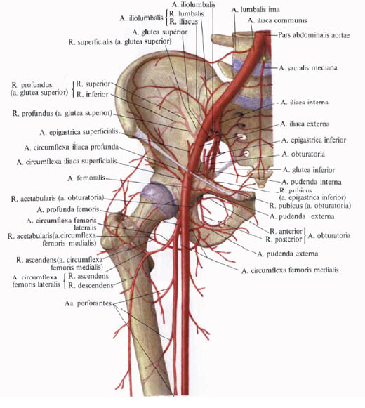
1) The medial artery surrounding the femur, a. Circumflexa femoris medialis, departs from the deep thigh artery behind the femoral artery, goes transversely inward and, penetrating between the ilio-lumbar and comb muscle into the muscle mass, leading the femur, bends the neck of the femur from the medial side.
From the medial artery, enveloping the femur, the following branches depart:
A) ascending branch, r. Ascendens, is a small stalk that goes up and down; Branched, approaches the comb muscle and the proximal part of the long adductor muscle;
B) transverse branch, r. Transversus, a thin stalk, directed downward and medially along the surface of the crested muscle, and, penetrating between it and the long adductor muscle, goes between the long and short adductor muscles; Blood supply to long and short adductor muscles, thin and outer blocking muscles;
C) deep branch, r. Profundus, - a larger trunk, which is a continuation of a. Circumflexa femoris medialis. It goes backwards, passes between the outer obturator muscle and the square muscle of the thigh, dividing itself here into the ascending and descending branches;
D) branch of the acetabulum, r. Acetabularis, is a thin artery, anastomosing with branches of other arteries, blood supplying the hip joint.
2) The lateral artery surrounding the femur, a, circumflexa femoris lateralis, a large trunk, extends from the outer wall of the deep thigh artery almost at its very beginning. Goes outside in front of the ilio-lumbar muscle, behind the sartorius muscle and the rectus muscle of the thigh; Going up to the large trochanter of the femur, is divided into branches:
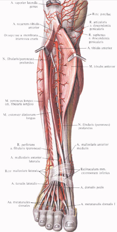
A) ascending branch, r. Ascendens, goes up and out, under the muscle stretching the broad fascia, and the middle gluteus muscle;
B) a descending branch, r. Descendens, more powerful than the previous one. It departs from the outer surface of the main trunk and lies under the straight muscle of the thigh, then descends along the furrow between the intermediate and lateral broad muscles of the thigh. Blood supply to these muscles; Reaching the region of the knee, anastomosing with the branches of the popliteal artery. On its way, it supplies blood to the head of the quadriceps muscle of the thigh and gives branches to the skin of the thigh;
C) transverse branch, r. Transversus, is a small stem, laterally guided; Blood supply to the proximal part of the rectus femur and lateral wide thigh muscle.
3) perforating arteries, aa. Perforantes, usually three, move away from the deep thigh artery at a different level and extend to the hamstring at the very line of attachment to the femur of the adductor muscles.
The first perforating artery begins at the level of the lower edge of the comb muscle; The second departs at the lower edge of the short adductor muscle and the third - below the long adductor muscle. All three branches perforate the adductor muscles at the place of their attachment to the femur and, coming out to the posterior surface, cut through the adductor, semimembranous, semitendinous muscles, the biceps femoris and the skin of this region.
The second and third perforating arteries give small branches to the femur - the artery feeding the thigh, aa. Nutriciae femaris.
4) Descending knee artery, a. Descendens genicularis, a rather long vessel, begins more often from the femoral artery in the leading canal, less often from the lateral artery circumscribing the femur. Going down, perforates together with the subcutaneous nerve, n. Saphenus, from the depth to the surface of the tendon plate, goes behind the sartorius muscle, bends around the inner condyle of the thigh and ends in the muscles of this region and the articular capsule of the knee joint.
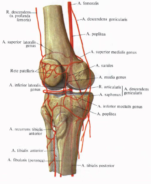
This artery gives the following branches:
A) the subcutaneous branch, r. Saphenus, in the thickness of the medial broad thigh muscle;
B) articular branches, rr. Articulares, taking part in the formation of the knee joint network, rete articulare genus, and patellar nets, rete patellae.


Comments
When commenting on, remember that the content and tone of your message can hurt the feelings of real people, show respect and tolerance to your interlocutors even if you do not share their opinion, your behavior in the conditions of freedom of expression and anonymity provided by the Internet, changes Not only virtual, but also the real world. All comments are hidden from the index, spam is controlled.