Posterior tibial artery
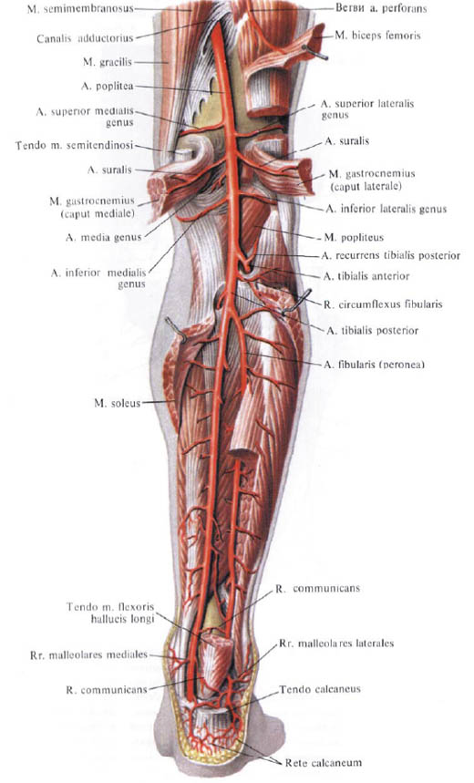
The posterior tibial artery , a.tibialis posterior, is a branch of the popliteal artery. It follows down the posterior surface of the tibia, lying behind the soleus muscle and in front of the posterior tibial muscle and the long flexor of the fingers. The artery is accompanied by two eponymous veins, and the lateral to it is the tibial nerve, tibialis.

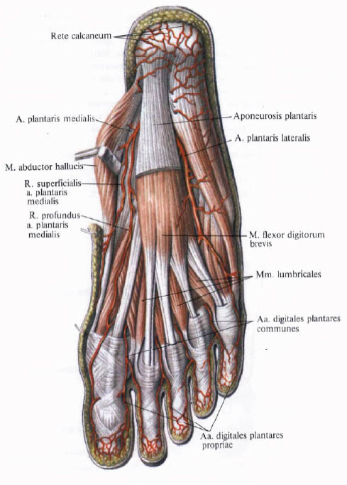
In the upper third of the leg from a. Tibialis posterior leaves a small stem entering the tibial nut and supplying it with blood, - the tibia feeding the tibia, a, nutricia tibiae. Going down and somewhat medial, the posterior tibial artery reaches the medial malleolus, which curves around the middle of the distance between it and the edge of the calcaneal tendon. Here, the artery is separated from the posterior edge of the medial ankle by the tendons of the posterior tibial muscle and the long flexor of the fingers and is located between the sheets of the flexor tendon holder separating it from the long flexor of the big toe. Having passed under retinaculum mm. Flexorum and further under the proximal site m. Abductor hallucis, the artery passes to the plantar surface of the foot and is divided here under the upper edge of the m. Abductor hallucis or else under retinaculum mm. Flexorum into two branches: the lateral plantar artery, and. Plantaris lateralis, and medial plantar artery, a. Plantaris medialis.

In its turn, the posterior brambler artery gives off a series of branches.
1. The artery surrounding the fibular bone, a. Circumftexus fibularu, departs from the main trunk at one hundredth of the beginning and is directed forward under the head of the fibula; Blood supply to the muscles of this area and takes part in the formation of the knee joint network.

2, Gonorabic artery, a. Fibularis (peronea), the largest branch of the posterior tibia. Begins from its initial department. Somewhat below the level of the head of the fibula is directed downward, laterally from the posterior tibial artery, near the fibula, along the posterior surface of the posterior tibial muscle, being covered behind (from the surface) by the long flexor of the big toe. At the level of the lateral ankle, the artery splits into the heel branches, rr. Calcanei, heading towards the ankle and to the heel net, rete calcaneum.
A number of branches leave the peroneal artery:
A) the perforating branch, r. Perforans, leaves 4 to 5 cm above the lateral ankle and, perforating the interosseous membrane, is directed downwards from the front of the shin; Here it anastomoses with the lateral anterior ankle artery, as well. Malleolaris anterior lateralis (from a. Tibialis anterior), taking part in the formation of the lateral ankle net, rete malleolare laterale, and calcaneal network, rete calcaneum;
B) lateral ankle branches, rr. Malleolares laterales, - small branches that enter the lateral ankle network. Anastomose with the anterior lateral ankle artery from the anterior tibial artery;
C) connecting branch, r. Communicans, a small trunk that extends at the ankle level medially along the posterior surface of the tibia and connects to a. Tibialis posterior.

3. Medial ankle branches, rr. Malleolares mediales, begin behind the medial malleolus and, heading forward, anastomosing with a. Malleolaris anterior medialis (from a. Tibialis anterior).
4. Heel branches, rr. Calcanei, only 2 - 4, are directed to the inner surface of the heel where, anastomosing with the lateral heel branches (from the peroneal artery), form a heel network.
5. The medial plantar artery. A. Plantaris medialia, leaving the retinaculum mm. Flexorum, goes along the medial edge of the plantar surface of the foot between m. Abductor hallucis and m. Flexor digitorum brevis, heading towards the I metatarsal bone. Passing between these muscles, the artery is divided into two branches - the surface and the deep:
A) surface branch, r. Superficialis, penetrates through m. Abductor hallucis, blood supply it and, going along the inner edge of the foot, reaches the first finger;
B) deep branch, r. Profundus, continues his course in the furrow between m. Abductor hallucis and m. Flexor digitorum brevis to the head I metatarsal bone, blood supply to the specified muscles and skin, anastomoses with a. Metatarsalis plantaris prima, and sometimes directly from the arcus plantaris.
6. Lateral plantar artery, a. Plantaris lateralis, larger in diameter than the previous one. Coming out from under m. Abductor hallucis, passes to the plantar surface of the foot, where between m. Flexor digitorum brevis and m. Quadratus plantae is directed slightly arcuate to the lateral edge of the foot. Here it goes forward and, reaching the base of the metatarsal V, gives its own plantar finger artery, a. Digitalis plantaris propria, to the lateral margin of the V finger, and turns to the medial side and passes between the deepest layer of the soles of the sole - mm. Interossei plantares and a more superficially positioned oblique head m. Adductor hallucis and tendons m. Flexor digitorum longus. Having thus passed in the medial direction, the artery forms a deep plantar arch, arcus plantaris profundus. Having reached the right interlacing interval, the arc joins r. Plantaris profundus (from a. Dorsalis pedis). Sometimes, between the lateral and medial plantar arteries under the plantar aponeurosis, at the level of the beginning of the tendons of the short flexor of the fingers, a surface plantar arc, arcus plantaris superficialis, is formed.
The following branches extend from the deep plantar arch:
A) plantar metatarsal arteries, aa. Metatarsales plantares, only four, are directed anteriorly in the spaces between the metatarsal bones. The distal ends of these arteries are called the common plantar digital artery, aa. Digitales plantares communes. At the level of the base of the first phalanges, each of them is divided into two own plantar finger arteries, aa. Digitales plantares propriae, which go along the edges of the fingers facing one another.
The first common plantar finger artery yields three own plantar finger arteries: one to the medial edge of the 2nd finger and two to the sides of the first finger:
B) a series of small branches to the muscles and bones of the plantar surface of the foot;
C) perforating branches, rr. Perforantes.


Comments
When commenting on, remember that the content and tone of your message can hurt the feelings of real people, show respect and tolerance to your interlocutors even if you do not share their opinion, your behavior in the conditions of freedom of expression and anonymity provided by the Internet, changes Not only virtual, but also the real world. All comments are hidden from the index, spam is controlled.