Anterior tibial artery
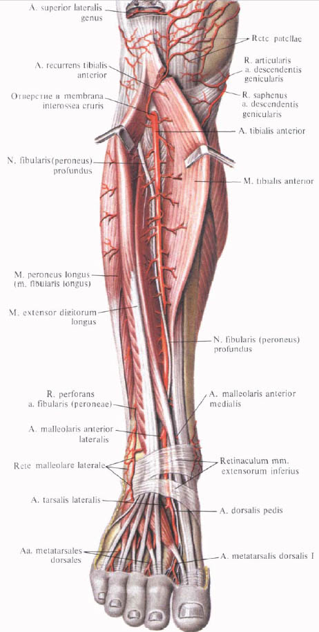
The anterior tibial artery , a.tibialis anterior, moving away from the popliteal artery, is directed forward, perforates in the proximal part of the interosseous membrane and leaves on the anterior surface of the tibia.

Here it goes along the anterior surface of the interosseous membrane, accompanied by two veins and a deep peroneal nerve, n. Peroneus profundus, which first follows laterally, and then crosses it and lies medially, going down. In the upper third of the tibia, the artery lies in the depth between m. Tibialis anterior and m. Extensor digitorum longus, and from the middle of the shin - between m. Tibialis anterior and m. Extensor hallucis longus.

In the distal part of the tibia, the artery is superficial and extends to the medial surface of the tibia, and at the ankle level lies on the capsule of the ankle, in the region of which, under the retinaculum mm. Extensorum inferius, passes on the rear surface of the foot, receiving the name of the dorsal artery of the foot, a. Dorsalis pedis.
On its way, the anterior tibial artery gives off a series of branches.
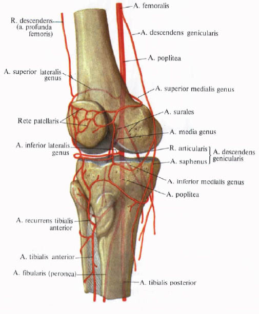
1. Posterior tibial return artery, a. Recurrens tibialis posterior, unstable, originates from the anterior tibial artery on the posterior surface of the tibia and is directed upward under the popliteal muscle, to the knee joint, takes part in the formation of the knee joint network.
2. Anterior tibial recurrent artery, a. Recurrens tibialis anterior, departs from the anterior tibial artery immediately after the latter passes through the interosseous membrane to the anterior surface of the tibia. It is directed upwards, penetrates the thickness of the anterior tibial muscle, passes to the anterior surface of the outer condyle of the tibia, anastomizing, like the previous artery, with the lateral and medial upper and lower knee arteries and branches of the middle knee artery; Takes part in the formation of the knee joint network.
3. Lateral anterior ankle artery, a. Malleolaris anterior lateralis, departs immediately proximal to the ankle joint; Goes under the tendon along the long extensor of fingers outside, on the anterior surface of the lateral ankle, where it participates in the formation of the lateral ankle net, rete malleolare laterale. On the way anastomoses with r. Perforans and rr. Malleolares laterales (from a. Fibularis), while giving a few small branches to the ankle joint.
4. Medial anterior ankle artery, a. Malleolaris anterior medialis, departs from the anterior tibial artery at the same level as the previous one. Going medially, passes under the tendon m. Tibialis anterior on the anterior surface of the medial malleolus and takes part in the formation of the ankle net.

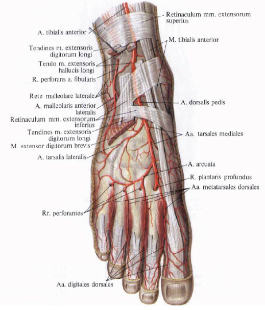
5. Posterior artery of the foot, a. Dorsalis pedis, is a continuation of the anterior tibial artery. Comes out from under retinaculum mm. Extensorum inferius and is directed forward along the rear of the foot, lying between m. Extensor hallucis longus and m. Etensor hallucis brevis. Having reached the interosteal interval between I and II metatarsal bones, it divides into a deep plantar artery, a. Plantaris profundus, and the first back metatarsal artery, a. Metatarsalis dorsalis prima.
The back artery of the foot gives a number of branches:
A) medial tarsal arteries, aa. Tarsales mediales, in the form of 2 - 3 small branches branch from the dorsal artery of the foot, go under the tendon m. Extensor hallucis longus to the medial margin of the foot, taking part in the formation of the medial malleolar network;
B) lateral tarsal artery, a. Tarsalis lateralis, the shore begins at the anterior end of the talus, goes laterally, and then forward along the tarsal bones under m. Extensor digitorum brevis and blood supply; Reaching the base of V metatarsal bone, anastomoses with arched artery, a. Arcuata;
C) an arcuate artery, a arcuata, begins at the proximal phalanx of II metatarsal bone, passes under m. Extensor digitorum brevis, directed forward and laterally, reaches the base of the metatarsal V, where it anastomizes with a. Tarsalis lateralis, forming an arterial arch. From the anterior periphery of the arched artery, II, III, IV posterior metatarsal arteries begin, aa. Metatarsales dorsales. They are straight, relatively thin vessels that follow forward, located in the three outer interosseous spaces on the rear interosseous muscles.
The initial sections of the rear metatarsal arteries II, III, IV at the level of the bases of the metatarsal bones through an interval between them are anastomosed by means of perforating branches, rr. Perforantes, with plantar metatarsal arteries, aa. Metatarsales plantares. At the level of the heads of metatarsal bones or somewhat distal, each posterior metatarsal artery divides into two rear finger arteries, aa. Digitales dorsales, which are directed anteriorly and lie along the edges of the back surface of the fingers facing each other.
The perforating artery between aa. Metatarsales dorsales and aa. Metatarsales plantares are poorly developed;
D) first dorsal metatarsal artery, a. Metatarsails dorsalis prima, - one of the two terminal branches of the dorsal artery of the foot. It goes in the first interosseous space along the back of the interosseous muscle, giving up three rear finger arteries, aa. Digitales dorsales: two to the first finger and one to the medial surface of the 2nd finger;
E) deep plantar artery, a. Plantaris profunda, is the second terminal branch of the dorsal artery of the foot. It perforates at the base of the first interosteal space m. Interosseus dorsalis prima and passes to the plantar surface of the foot, anastomosing with the terminal part of the lateral plantar artery from the posterior tibial artery; Forms a plantar arch.
Arterial nets. On the lower limb there is a series of anastomoses between the large arterial trunks and their branches, which, especially in the joints, form arterial nets.
1. The knee articular network, rete articulare genus, is a dense arterial network in the formation of which branches are involved, departing from: a) a. Descendens genicularis (from A. femoralis); B) a. Superior medialis genus, a. Superior lateralis genus, a. Media genus, a. Inferior medialis genus, a. Inferior lateralis genus (all from a. Poplitea); C) r. Circumflexus fibularis (from a. Tibialis posterior); D) a. Recurrens tibialis posterior (from a. Tibialis anterior); E) a. Recurrens tibialis anterior (from a. Tibialis anterior).
2. The medial malleolar network, rete malleolare mediale, is formed by the following branches: a) rr. Malleolares mediales (from a. Tibialis anterior); B) a. Malleolaris anterior medialis (from a. Tibialis anterior); C) aa. Tarsales mediales (from a. Dorsalis pedis).
3. The lateral ankle net, rete malleolare laterale, is formed by the following branches: a) rr. Malleolares laterales from a. Fibularis (peronea); B) branches from r. Perforans a. Fibularis (peronea); C) a. Malleolaris anterior medialis (from a.tibialis posterior); D) back branches a. Tarsalis lateralis (from a. Dorsalis pedis).
4. The calcaneal network, rete calcaneum, lies on the posterior surface of the calcaneus calcaneus. The formation of this network involves: a) rr. Calcanei from a. Tibialis posterior; B) rr. Calcanei from a. Fibularis (peronea).
5. Anastomoses of the arteries of the plantar and dorsal surface foot are described earlier.


Comments
When commenting on, remember that the content and tone of your message can hurt the feelings of real people, show respect and tolerance to your interlocutors even if you do not share their opinion, your behavior in the conditions of freedom of expression and anonymity provided by the Internet, changes Not only virtual, but also the real world. All comments are hidden from the index, spam is controlled.