Breast-and-groove joints
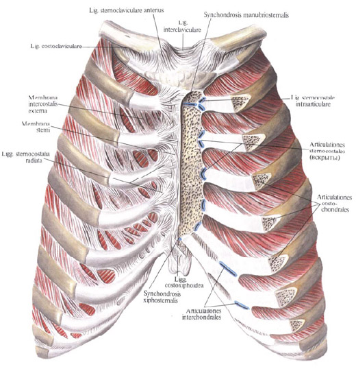
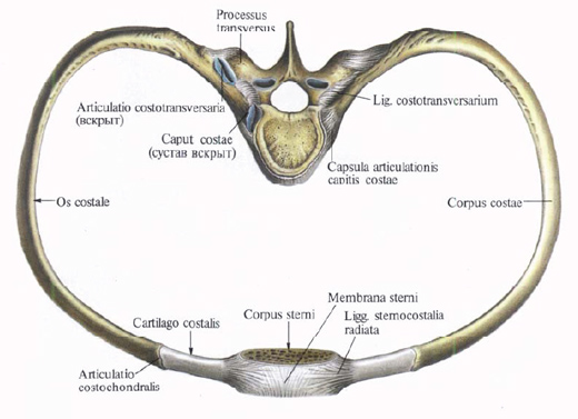
Chest-rib joints . The anterior ends of the ribs end with costal cartilages. The bony part of the ribs connects to the reticular cartilages by the costal-cartilaginous joints, articutationes costochondrales, and the periosteum of the rib extends into the supracriphon of the corresponding costal cartilage, and the connection between them is impregnated with age with age.

The costal cartilage of the 1st rib fuses with the sternum. The costal cartilages of the II-VII ribs are articulated with rib sternum incisions, forming the sternocostal joints, articulationes sternocostales.

The cavity of these joints represents a narrow, vertically arranged gap, which in the cavity of the joint of the 2nd costal cartilage has an intraarticular sternocostal ligament, lig. Sternocostal intraarticulare. It goes from the costal cartilage of the 2nd rib to the joint of the arm and the body of the sternum.
In the cavities of other sternocostal joints this ligament is weakly expressed or absent.
The joint capsules of these joints, formed by the perichondrium of the costal cartilage, are strengthened by radial sternocostal ligaments, ligg. Sternocostalia radiata, of which the anterior, more powerful than the posterior ones. These ligaments go radiantly from the end of the costal cartilage to the anterior and posterior surfaces of the sternum, forming crossings and bindings with the same ligaments of the opposite side, as well as with the underlying and underlying ligaments. As a result, a sturdy fibrous layer covering the sternum is formed - the sternum membrane, the membra na sterni.
Fiber bundles that follow from the front surface of the VII VII costal cartilage obliquely downward and medially to the xiphoid process form rib-xipedal ligaments, ligg. Costoxiphoidea.
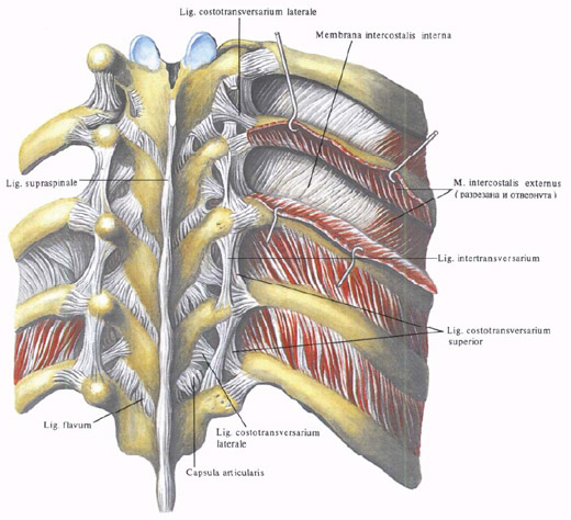
In addition, in the intercostal spaces are located the outer and inner intercostal membranes.

The outer intercostal membrane, the tetra intercostalis externa, lies on the front surface of the thorax in the area of the costal cartilage. The bundles forming it start from the lower edge of the cartilage and, running obliquely down and anteriorly, terminate at the upper edge of the underlying cartilage. The internal intercostal membrane, membrana intercostalis interna, is located in the posterior sections of the intercostal spaces. Its tufts start from the upper edge of the rib and, moving obliquely upward and anteriorly, attach to the lower edge of the overlying rib. There are no intercostal muscles in the locations of the membranes. Both membranes strengthen the intercostal space.
The costal cartilages from V to IX ribs are interconnected by dense fibrous tissue and interchilar joints, arliculationes interchondrales. The tenth rib is connected by a fibrous tissue to the cartilage of the IX rib, and the cartilages of the XI and XII ribs terminate freely between the abdominal muscles.


Comments
When commenting on, remember that the content and tone of your message can hurt the feelings of real people, show respect and tolerance to your interlocutors even if you do not share their opinion, your behavior in the conditions of freedom of expression and anonymity provided by the Internet, changes Not only virtual, but also the real world. All comments are hidden from the index, spam is controlled.