Temporomandibular joint
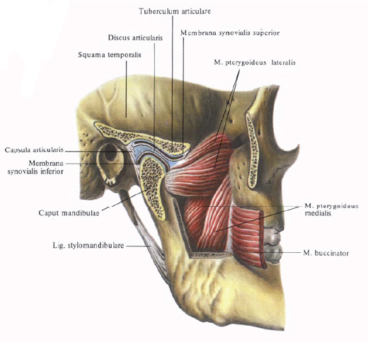
Temporomandibular joint , artculatio temporomandibularis, paired. It is formed by the head of the mandible, caput mandibulae in the mandibular fossa, fossa mandibularis, and articular tuberculum, tuberculum articulare, scaly part of the temporal bone. The mandibular heads are cylindrical; Long converging axes with their continuation converge at an obtuse angle at the anterior edge of the large occipital orifice.
The mandibular fossa of the temporal bone does not fully enter the temporomandibular joint cavity.

It distinguishes two parts: the extra-capsular part of the mandibular fossa that lies behind the stony-scaly slit, and the intracapsular part of the mandibular fossa is anterior to it. This part of the fossa is enclosed in a capsule, which extends to the articular tubercle, reaching its anterior margin. The articular surfaces are covered with connective tissue cartilage. In the joint cavity lies biconcave, oval-shaped fibrous cartilaginous plate - articular disc, discus articularis.

Located in the horizontal plane, the disk with its upper surface lies with the articular tubercle, and the lower one with the head of the lower jaw. It fuses around the circumference with the joint capsule and divides the joint cavity into two parts that do not communicate with each other - the upper and lower ones. The cavity of each department respectively lining the upper synovial membrane, membrana synovialis superior, and the lower synovial membrane, membrana synovialis inferior. To the inner edge of the disc is attached a part of the tendon bundles of the lateral pterygoid muscle, m. Pterygoideus lateralis.
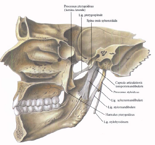
The joint capsule is attached along the edge of the articular cartilage; On the temporal bone it is fixed in front along the anterior slope of the articular tubercle, posteriorly along the anterior margin of the stony-tympanic gap, laterally at the base of the zygomatic process, medially reaching the spine of the sphenoid bone; On the lower jaw articular capsule covers her neck, attaching to her behind a bit lower than the front.

To the ligaments of the temporomandibular joint are:
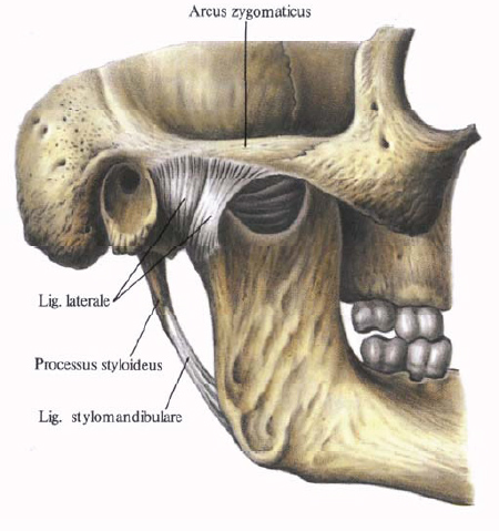
1. Lateral ligament, tig. Laterale, begins from the base of the zygomatic process and is directed to the outer and posterior surfaces of the neck of the lower jaw. Some of the bundles of this ligament are weaved into the capsule of the joint. In a bunch of distinguished
Front and back.
2. Medial ligament, lig. Mediale, passes along the ventral surface of the capsule of the temporomandibular joint. It originates from the inner edge of the articular surface and the base of the spine of the sphenoid bone and is attached to the posterior-inner surface of the neck of the articular process.
In addition, there are ligaments related to the temporomandibular joint, but not associated with the joint capsule: wedge-mandibular ligament, tig. Sphenomandibulare, begins from the spine of the sphenoid bone and is attached to the tongue of the lower jaw; Ankylogenous ligament, lig. Stylomandibulare, is directed from the styloid process to the angle of the lower jaw. Temporomandibular joint refers to the type of block joints. When moving in the joint, it is possible to lower and raise the lower jaw, push it forward and return to its original position, shift left and right. Such a variety of movements in the block joint is due to a combination of movements of the joint head simultaneously in the right and left joints, as well as the presence of a fibrous joint disk in each joint dividing the joint cavity into the upper and lower divisions, which allows diversifying the movements of the lower jaw. For example, lowering the lower jaw occurs when the head of the lower jaw rotates around the horizontal axis under the articular disc, i.e., in the lower part of the joint cavity; Extension of the jaw forward occurs when the head of the jaw moves together with the disc on the articular tubercle, i.e. In the upper part of the cavity. Movement of the lower jaw to the sides is also performed with the participation of the joint disc, and in the joint on the side of displacement (in the right when moving to the right and vice versa), the head of the lower jaw rotates under the articular disc, and in the opposite joint - the head extends to the articular tubercle above the disc.


Comments
When commenting on, remember that the content and tone of your message can hurt the feelings of real people, show respect and tolerance to your interlocutors even if you do not share their opinion, your behavior in the conditions of freedom of expression and anonymity provided by the Internet, changes Not only virtual, but also the real world. All comments are hidden from the index, spam is controlled.