


Elbow joint. Elbow joint structure
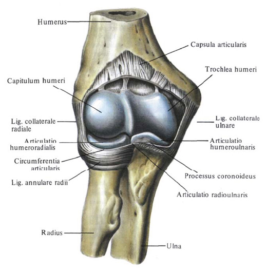
The elbow joint , articulatio cubiti, is formed by the articular surface of the distal epiphysis of the humerus - its block and head of the condyle, articular surfaces on the ulnar bone - with blocky and radial incisions of the ulna, as well as the head and articular circumference of the radius . The joint is complex (articulatio composita), since it consists of three joints, each of which has its own form.
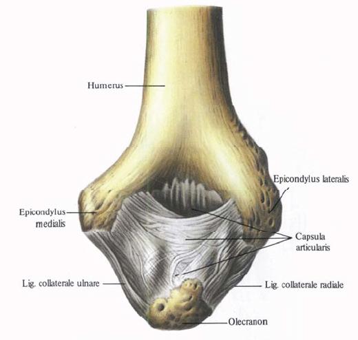
The structure of the elbow joint.
In the elbow joint, flexion and extension, pronation and supination are possible. The articular surfaces of the bones forming the joints are covered with hyaline cartilage.
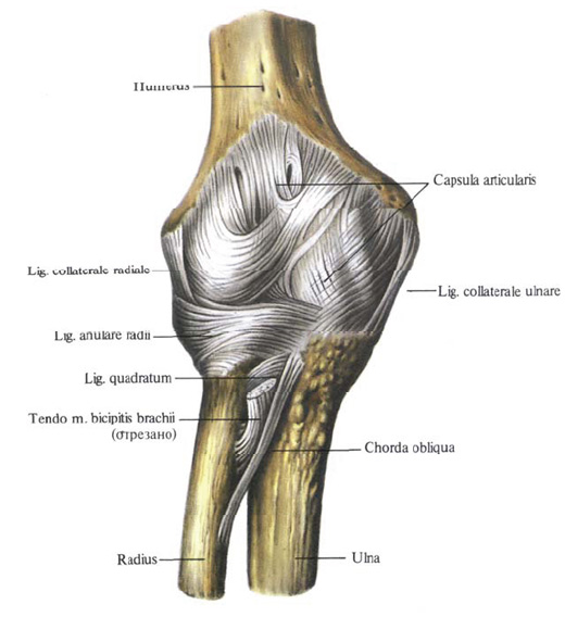
The joint capsule surrounds all three joints. On the humerus, it is fixed in front of the edge of the coronary and radial pits, along the sides - along the periphery of the bases of the epicondyle (leaving them free), almost at the edge of the articular surface of the block and head of the condyle of the humerus, and behind - slightly below the upper margin of the pit of the ulnar process. On the ulna, the joint capsule is attached along the edge of the block and radial scissors, and on the radius bone to the neck of the radius, forming a saccular protrusion here. The joint capsule in the anterior and posterior parts of the joint is thin and slightly stretched, and in the lateral it is strengthened with ligaments. Its synovial membrane also covers those parts of the bones that are in the joint cavity, but are not covered with cartilage (neck of the radial bone, etc.).
In the cavity of the elbow joint, three joints are distinguished: the mediastinal, the brachial and the proximal radiolucent.
1. The articulate joint, articulutio humeroulnaris, is located between the surface of the humerus block and the blocky ulnar bone cutting. It refers to uniaxial and is a block-shaped joint, with a helical deflection of the articular surfaces.
2. The brachium joint, articulatio humeroradialis, is formed by the head of the condyle of the humerus and the joint fossa on the head of the radius, refers to the globular joints, in spite of the fact that in fact the motions in it are made around not three but only two axes-the frontal and vertical axes.
3. The proximal radioclavicular joint, articulatio radioulnaris proximalis, lies between the radial incision of the ulna and the articular circumference of the radial head: it is a typical cylindrical joint with rotation around one vertical axis.
In the brachial joint, flexion and extension are possible, which occur simultaneously with the movement of the radial bone in the brachial joint. In this joint, it is also possible to rotate the radius along its long axis inwards and outwards. In addition, in the proximal radiophila joint, rotation of the radial bone occurs with simultaneous movement in the shoulder joint.
The following ligaments are connected to the elbow joint:
1. Elbow collateral ligament, lig. Collaterale ulnare, goes from the base of the medial epicondyle of the humerus downwards and, fan-like expanding, is attached at the edge of the block-shaped notch of the ulna.
2. Radial collateral ligament, lig. Collaterale radiale, starts from the base of the lateral padmichele of the humerus, down to the outer surface of the radial head, where it divides into two bundles. These bundles take a horizontal direction and, bending the head of the radial bone in front and behind, attach to the edges of the radial cutting of the ulna. The superficial layers of the ligament are fused with the extensor tendons. The deep pass into the annular bundle of the radius.
3. Ring bundle of radius, lig. Anulare radii, covers the joint circumference of the radial head from the anterior, posterior and lateral sides, and, attached to the anterior and posterior edges of the radial incision of the ulna, holds the radius of the ulnar bone.
4. Square bundle, lig. Quadratum is a bundle of fibers that connect the distal edge of the radial cutting of the ulna with the neck of the radius.
In the elbow joint lateral movements are absent, since they are inhibited by strong collateral ligaments. In general, the elbow joint is a block-shaped joint with a somewhat helical shape of the sliding of the joint surfaces.
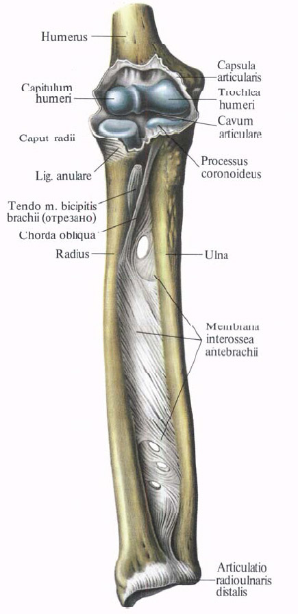
In addition to the annular ligament of the radius, in the fixation of the bones of the forearm takes part the interosseous membrane of the forearm.
The interosseous membrane of the forearm, membrana interossea antebrachii, fills the gap between the radial and ulna bones, attaching to their interosseous edges and forming radiolucent syndesmosis, syndesmosis radioulnaris.
It is formed by strong fibrous tufts, which go obliquely from top to bottom from the radial bone to the ulnar bone. One of these beams has the opposite direction: it follows from the tuberosity of the ulna to the tuberosity of the radius and is called the chorda chorda, chorda obliqua. The membrane has holes through which the vessels and nerve pass. A series of muscles of the forearm begins from her palmar and dorsal surfaces.

Comments
When commenting on, remember that the content and tone of your message can hurt the feelings of real people, show respect and tolerance to your interlocutors even if you do not share their opinion, your behavior in the conditions of freedom of expression and anonymity provided by the Internet, changes Not only virtual, but also the real world. All comments are hidden from the index, spam is controlled.