The sacroiliac joint
The sacroiliac joint , articulatio sacroiliaca, is a paired joint, is formed by ileal bones and sacrum.

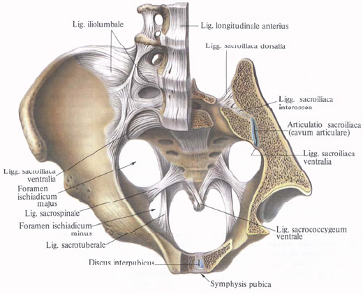
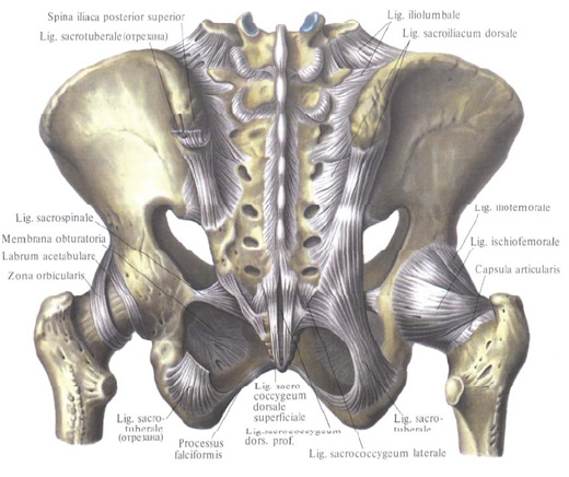
Articular anatomical surfaces, facies auriculares, iliac bones and sacrum are flat, covered with fibrous cartilage, Joint capsule is attached along the edge of articular surfaces and tightly stretched. The ligamentous device is represented by strong, strongly stretched fibrous bundles, located on the front and back surfaces of the joint. On the front surface of the joint are the anterior sacroiliac ligament, ligg. Sacroiliaca anteriora (ventralia). They are short bundles of fibers that extend from the pelvic surface of the sacrum to the ilium.
On the back surface of the joint are several ligaments:
1. Mezhostnye sacroiliac ligaments, ligg. Sacroiliaca interossea, lie behind the sacroiliac joint, in the interval between the bones forming it, attaching at their ends to the iliac and sacral tuberosity.
2. Posterior sacroiliac ligaments, ligg. Sacroiliaca posteriora (dorsalia). Individual bundles of ligament ethics, starting from the inferior posterior iliac spine, attach to the lateral sacral ridge at level II-III sacral orifices. Others follow from the upper posterior iliac awn downward and somewhat medially, attaching to the posterior surface of the sacrum in the region of the IV sacral vertebra.
The sacroiliac joint belongs to the inferior joints.
The pelvic bone, except for the sacroiliac joint, is connected to the vertebral column by a series of powerful ligaments, which include the following:
1. Sacrum-ligamentous ligament, lig. Sacrotuberale, begins from the medial surface of the ischial hillock, and, going upward and medially, expands in a fan-shaped manner; Is attached to the outer edge of the sacrum and coccyx. Part of the fibers of this ligament passes to the lower part of the branch of the ischium and, continuing along it, forms a sickle-shaped process, porcessus falciformis.

2. Sacrum-awned ligament, lig. Sacrospinale, begins from the ischium awn, goes medially and posteriorly and, located in front of the previous ligament, is attached along the rim of the sacrum and partly the coccyx.
Both ligaments, together with large and small sciatic cuttings, limit two holes: a large sciatic, foramen ischiadicum manus, and a small ischial, foramen ischiadicum minus. Through these holes pass out of the pelvis muscles, as well as blood vessels and nerves.
3. Ilio-lumbar ligament, lig. Iliolumbale, begins from the anterior surface of the transverse processes of the IV and V lumbar vertebrae, is directed to the outside and is attached to the posterior sections of the iliac crest and the medial surface of the ileal wing. This ligament strengthens the lumbosacral joint, articulatio lumbosacralis.


Comments
When commenting on, remember that the content and tone of your message can hurt the feelings of real people, show respect and tolerance to your interlocutors even if you do not share their opinion, your behavior in the conditions of freedom of expression and anonymity provided by the Internet, changes Not only virtual, but also the real world. All comments are hidden from the index, spam is controlled.