Ankle joint
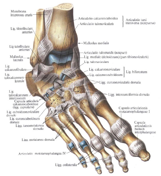
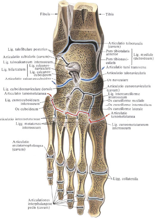
The ankle joint, articulatio talocruralis, is formed by the articular surfaces of the distal epiphyses of the tibial and fibular bones and the articular surface of the talus block. On the tibia the articular surface is represented by the lower articular surface of the tibia, facies articularis inferior tibiae, and the articular surface of the ankle, facies articularis maleoli. On the fibula also there is an articular surface of the ankle, faciei articularis maleoli fibulae.

The articular surface of the talus bone from above has the shape of a block, and on the sides it is represented by flat articular sites - lateral and medial ankle surfaces, facies malleolares lateralis et medialis. The shank bones in the form of a fork cover the block of the talus bone.
The joint capsule is attached to the margin of the articular cartilage for a long time and only a few steps away from it on the anterior surface of the body of the talus, attaching itself to the neck of the talus. The anterior and posterior parts of the articular capsule are slightly strained.
Ligaments of the ankle joint lie on its lateral surfaces.
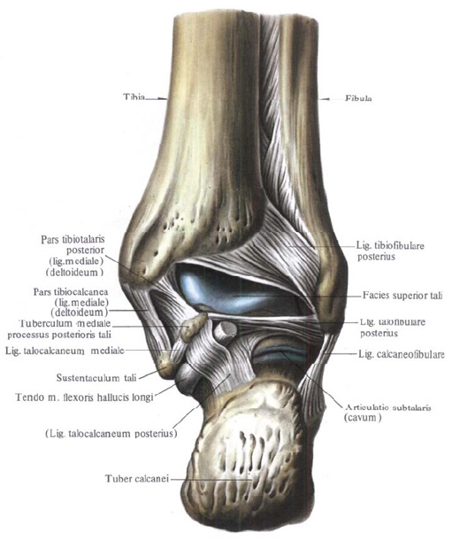
1. Medial (deltoid) ligament, lig. Mediale (deltoideum), which includes the following parts:

A) the anterior tibial part, pars tibiotalaris anterior, extends from the anterior edge of the medial ankle downward and forward and is attached to the posterior medial surface of the talus.

B) the tibial-navicular part, pars tibionavicularis, longer than the previous one, starts from the medial malleolus and reaches the rear surface of the scaphoid bone;

C) tibial calcified part, pars tibiocalcanea, taut between the medial ankle and the support of the talus;
D) the posterior tibial part, pars tibiorularis posterior, extends from the posterior margin of the medial ankle downward and laterally and is attached to the posterior medial parts of the body of the talus.
2. Anterior collateral ligament, lig. Talofibulare anterius, from the anterior edge of the lateral ankle to the lateral surface of the collar of the talus.
3. The heel-peroneal ligament, lig. Calcaneofibulare, starts from the outer surface of the lateral malleolus, is directed downward and backward and attached to the lateral surface of the calcaneus.

4. Posterior incurvulatory ligament. Lig. Talofibulaere posterius, goes from the posterior edge of the lateral ankle almost horizontally to the lateral tubercle of the posterior process of the talus.
The ankle joint is a block-shaped joint, It can have a helical movement.


Comments
When commenting on, remember that the content and tone of your message can hurt the feelings of real people, show respect and tolerance to your interlocutors even if you do not share their opinion, your behavior in the conditions of freedom of expression and anonymity provided by the Internet, changes Not only virtual, but also the real world. All comments are hidden from the index, spam is controlled.