


Knee-joint
In the formation of the knee joint, articutatio genus, three bones: the distal epiphysis of the femur, the proximal epiphysis of the tibia and the patella. The joint surface of the condyles of the femur is ellipsoidal, the curvature of the medial condyle is greater than that of the lateral one. On the anterior surface of the bone, between the condyles, is the patellar surface, facies patellaris. With a small vertical groove, this surface is divided into a smaller medial and larger lateral areas that articulate with the corresponding articular surfaces located on the posterior articular surface of the patella, facies articularis. The upper articular surfaces of the condyles of the tibia are slightly concave and do not correspond to the curvature of the articular surfaces of the condyles of the femur. This discrepancy somewhat aligns between the condyles of the femoral and tibia bones interarticular cartilages - the medial and lateral menisci, menisci mediatis et lateralis. Menisci are trihedral cartilaginous plates. The outer margin is thickened and fused with the joint capsule; Internal, free, the edge is pointed and turned into the joint cavity. The upper surface of the meniscus is concave, the lower surface is flattened. The outer edge of the meniscus almost repeats the configuration of the upper edge of the tibial condyles (therefore the lateral meniscus resembles a part of the circumference, and the medial has a semilunar shape). Menisci attached to the front and back to the intercondylar elevation of the tibia. The front edges of both menisci are connected by a transverse ligament of the knee, lig. Transversum genus.
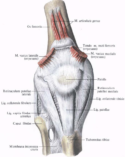
The joint capsule is attached to the edges of the femoral, tibial bones and to the patella. On the femur, it is attached under the epicondyle, so that they remain outside the cavity; In front and behind the capsule rises approximately 1 cm above the joint surface. On the tibia, the capsule is fixed along the edge of the joint surface, and is attached to the patella along its articular surface in such a way that the anterior surface of the patella is outside the joint cavity. The synovial membrane lining the articulating surfaces of bones to the line of articular cartilage. Going into the cavity of the joint, it surrounds the cruciate ligaments, forming numerous synovial villi, villi synoviales, and synovial folds, plicae synoviales. The most developed folds of the synovial membrane are: pterygoid folds, plicae alares, which run along the sides of the patella in the direction of its apex and contain a podadikolennik fat body between their leaves, corpus adiposum infrapatellare; Synaptic synovial fold, plica synovialis infrapatellaris; Lying below the patella and representing the continuation of the pterygoid folds. It begins at the apex of the patella, goes into the cavity of the knee joint and is attached to the anterior margin of the intercondylar fossa of the femur.
The knee joint capsule forms a series of synovial bags, bursae synoviales, lying along the muscles and tendons, but not communicating with the joint cavity. The largest protrusion of the articular capsule is the supra-nasal lining bag, bursa suprapatellaris. It is located above the patella, between the tendon of the quadriceps muscle and the femur; Sometimes it can be isolated.
The ligaments of the knee joint are divided into two groups: ligaments located outside the joint cavity (extra-capsular ligaments), and ligaments lying inside the joint capsule (intracapsular ligaments). On the lateral surfaces of the joint, there are the following well developed lateral ligaments.
1. Tibial collateral ligament, lig. Collaterale tibiale, follows from the medial epicondyle of the femur down, along the way it fuses with the capsule of the joint and the medial meniscus, reaching the upper epiphysis of the tibia.
2. Cholera collateral ligament, lig. Collateral fibular, which is already the previous one, starts from the lateral epicondyle of the thigh, goes down, like the previous one, and gives a number of its bundles to the joint capsule and is attached to the outer surface of the fibular head.
The anterior sections of the joint capsule are strengthened with ligaments that are directly related to the tendon of the quadriceps femoris muscle. The tendon of this muscle approaches the patella, covers it from all sides and continues downward, reaching the tibia. Most of the bundles coming from the apex and adjacent surfaces of the patella reach the tuberosity of the tibia. This cord is called the patella ligament, lig. Patellae. The lateral parts of the tendon bundles of this ligament extend from the patella to the outer and inner condyles of the tibia, forming respectively a lateral supporting patellar patella, retinaculum patellae laterale, and a medial supporting patellar patella, retinaculum patellae mediale.
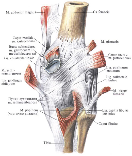
In the supporting patella ligaments, there are also horizontal bundles that attach to the epicondyle of the femur. Supporting ligaments of the patella play an important role in movements in the joint, holding the patella in the desired position. The posterior parts of the joint capsule are strengthened with an oblique popliteal ligament, lig. Popliteum obliquum, which is part of the bundles of the tendon of the semimembranous muscle, t. Semimembranosis. The ligament follows from the medial condyle of the tibia to the lateral condyle of the femur and part of its bundles are weaved into the joint capsule.
In addition to this ligament, an arcuate popliteal ligament is constantly present in this section of the articular capsule, lig. Roleritis of the arcuatum, which starts from the lateral condyle of the femur and the head of the fibula and is attached in the middle sections to the lig. Poplitei obliqui and further to the outer condyle of the tibia.
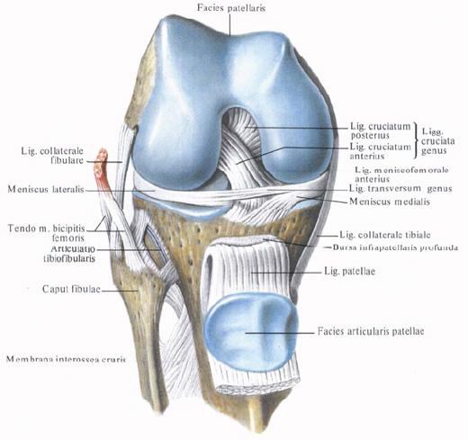
Inside the cavity of the knee joint are the cruciform ligaments of the knee, ligg. Cruciata genus, which are divided into:
A) anterior cruciate ligament, lig. Cruciatum anterius, starting from the inner surface of the lateral condyle of the thigh and following forward and medially; Is attached to the anterior intercondylar field, area intercondylaris anterior, tibia;
B) posterior cruciate ligament, lig. Cruciatum posterius, which begins on the inner surface of the medial condyle of the hip, and then, going back and medially, crosses with the anterior cruciate ligament and is attached to the posterior intercondylar field of the tibia.
In addition, there are three more ligaments directly related to the meniscus:
1. The transverse ligament of the knee, lig. Transversum genus, connects the anterior surface of both menisci.
2. Anterior meningoconical ligament, lig. Meniscofemorale anterius, starts from the anterior section of the medial meniscus, goes upward and lateral to the medial surface of the lateral condyle of the thigh.
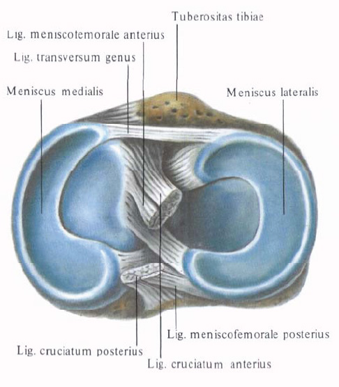
3. Back menisci ligament, lig. Meniscofemorale posterius, follows from the posterior edge of the lateral meniscus upward and medially to the inner surface of the medial condyle of the thigh.
The knee joint is a condylar joint, and in its unfolded position it works like a block-shaped joint. When the shank is bent, a rotational motion occurs in it.

Comments
When commenting on, remember that the content and tone of your message can hurt the feelings of real people, show respect and tolerance to your interlocutors even if you do not share their opinion, your behavior in the conditions of freedom of expression and anonymity provided by the Internet, changes Not only virtual, but also the real world. All comments are hidden from the index, spam is controlled.