


Median atlanto-joint
The medial atlanto-axial joint , articulatio atlanto-axialis mediana, is formed between the posterior surface of the anterior arch of the atlas (fovea dent is) and the tooth of the axial vertebra. In addition, the posterior articular surface of the tooth forms a joint with a transverse ligament of the atlas, lig transversum atlantis.
Joints of the tooth belong to the group of cylindrical joints. In them, it is possible to rotate the atlas together with the head around the vertical axis of the axial vertebra tooth, ie, turn the head to the right and to the left.
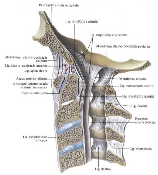
The ligamentous apparatus of the medial atlanto-osseous joint includes:
1. Cover membrane, membrana tectoria, which is a broad, fairly dense fibrous plate, stretched from the anterior edge of the large occipital opening to the body of the axial vertebra. This membrane is called the integumentary, because it covers the posterior (from the side of the spinal canal) tooth, the transverse bunch of the atlas and other formations of this joint. It is considered as part of the posterior longitudinal ligament of the vertebral column.
2. The cruciate ligament of the atlas, lig. Cruciforme atlantis, consists of two beams - longitudinal and transverse. The transverse bundle is a tight connective tissue cord stretched between the inner surfaces of the lateral mass of the atlas. It is attached to the posterior joint surface of the tooth of the axial vertebra and strengthens it. This beam is called the transverse bunch of the atlant, lig. Iransversum atlantis. Longitudinal fascicles, fasciculi longitudinales, consist of two, upper and lower, legs. The upper leg extends from the middle part of the transverse ligament of the atlas and reaches the anterior surface of the large occipital foramen. The lower leg, which also starts from the middle part of the transverse ligament, is directed downward and attached to the posterior surface of the body of the axial vertebra.

3. Ligament of the apex of the tooth, lig. Apicis dentis, stretches between the tip of the tooth of the axial vertebra and the middle part of the anterior margin of the large occipital opening. This ligament is considered as a rudiment of the dorsal string (chorda).
4. Pterygoid ligaments, ligg. Alaria, are formed by bundles of connective tissue fibers stretched between the lateral surfaces of the tooth of the axial vertebra and the internal surfaces of the occipital condyles, condyli occipitales.
You will be interested to read this:

Comments
When commenting on, remember that the content and tone of your message can hurt the feelings of real people, show respect and tolerance to your interlocutors even if you do not share their opinion, your behavior in the conditions of freedom of expression and anonymity provided by the Internet, changes Not only virtual, but also the real world. All comments are hidden from the index, spam is controlled.