


Internal iliac artery
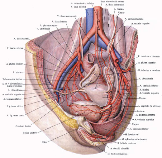
Internal iliac artery , a. Iliaca interna, departs from the common iliac artery and is directed downward into the cavity of the small pelvis, located along the line of the sacroiliac joint. At the upper end of the large sciatic foramen is divided into the anterior and posterior trunks. Branches that depart from these trunks are directed to the walls and organs of the small pelvis and are therefore divided into the internal and parietal.
Internal branches
1. Umbilical artery, a. Umbilicalis, in the embryonic period - one of the largest branches of the internal iliac artery. It departs from the anterior trunk of the latter and, moving forward along the lateral wall of the pelvis, goes to the side wall of the bladder, and then under the peritoneum it goes along the back surface of the anterior wall of the abdominal cavity up to the navel area. Here, together with the vessel of the opposite side of the same name, the umbilical artery is part of the umbilical cord. After birth, the lumen of the vessel is closed for a considerable length (obliterated part, pars occlusa), and the artery becomes a medial umbilical ligament. The initial section of the vessel remains passable - this is an open part, pars patens, which functions throughout life. It leaves the following arteries:
A) upper arteries, aa. Vesicales superiores, only 2 - 4, depart from the initial part of the umbilical artery. They are directed to the upper parts of the bladder and supply the top of the bladder;
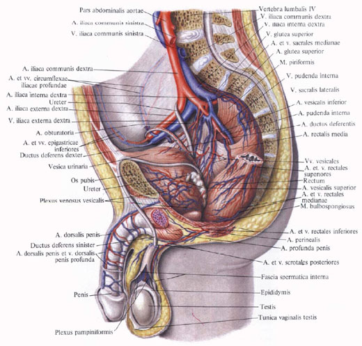
B) artery of the vas deferens, a. Ductus deferentis, goes forward and, reaching the vas deferens, divides into two branches that follow along the duct. One of them, together with the duct, enters the spermatic cord, anastomosing with a. Testicularis. Together with the spermatic cord passes through the inguinal canal and reaches the epididymis. Another branch goes along with the vas deferens to the seminal vesicles. From it in this area the ureteral branches depart, rr. Ureterici, to the pelvic portion of the ureter.
2. Lower colibuty artery, a. Vesicalis inferior, departs from the internal iliac artery and, approaching the bottom of the bladder, anastomizes with the branches of the upper urinary artery. It gives the representative branches, rr. Prostatici, and in women - fickle branches to the vagina.
3. Uterine artery, a. Uterina (corresponds to the artery of the vas deferens in men), departs from the anterior trunk of the internal iliac artery and, located under the peritoneum, goes forward and medially at the base of the broad ligament, reaching the lateral wall of the uterus at the level of its neck; On the way crosses deeper than the ureter. Going to the wall of the uterus, gives the descending vaginal branches, rr. Vaginales that go along the anterolateral wall of the vagina, giving him branches that anastomose with the same branches of the opposite side. The uterine artery rises along the side wall of the uterus to the corresponding horn of the uterus, where it sends the scroll branches, rr. Helicini. The artery is anastomosed with the ovarian artery (branch of the abdominal part of the aorta) and gives off the tubal branches, rr. Tubarii, to the fallopian tube and ovarian branches, rr. Ovarici, to the ovary.
4. The middle rectum artery, a. Rectalis media, - a small vessel, sometimes absent. It starts from the anterior trunk of the internal iliac artery, usually alone, but sometimes from the lower columbar artery or internal genital artery, a. Pudenda interna; Blood supply to the middle part of the rectum. From the artery a number of small branches branch to the prostate gland and seminal vesicles. In the rectal wall, the artery anastomoses with the upper (branch of the inferior mesenteric artery) and the lower arteries of the rectum, a. Rectalis superior et a. Rectalis inferior.
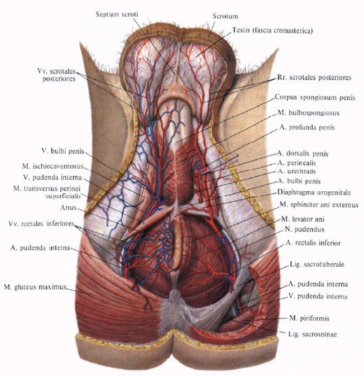
5. Internal sexual artery. A. Pudenda interna, departs from the anterior trunk of the internal iliac artery, goes down and outward and emerges from the small pelvis through the sub-tubus orifice. Then he goes around the sciatic wasteland and, moving medially and forward, again enters the cavity of the small pelvis through a small sciatic hole, already below the pelvic diaphragm, getting into the ischial-anal fossa. Following the lateral wall of this fossa, the internal sexual artery reaches the posterior margin of the urogenital diaphragm. Heading anteriorly along the lower branch of the pubic bone, at the edge of the transverse transverse perineal muscle, the artery perforates from the depth to the surface of the urogenital diaphragm and divides into a series of terminal branches:
A) dorsal artery of the penis (clitoris), a. Dorsalis penis (clitoridis), is essentially a continuation of a. Pudenda interna. Together with the same-named artery of the opposite side passes along the sinewy ligament of the penis, on the sides of the middle dorsal member of the deep dorsal vein of the penis, v. Dorsalis penis profunda, to its head, giving branches to the scrotum and cavernous bodies;
B) the artery of the bulb of the penis, a. Bulbi penis, [in women - artery bulbs of the vestibule (vagina), a. Bulbi vestibuli (vaginae)], blood supply to the bulb of the penis, bulbous-spongy muscle, the mucosa of the posterior part of the urethra and bulbourethral glands;
C) the urethral artery, a. Urethralis, enters the spongy body of the urethra and follows it to the glans penis, where it anastomizes with a. Profunda penis. In women it ends with two branches: to the urethra and to the bulb of the vestibule;
D) deep artery of the penis (clitoris), a. Profunda penis (clitoridis), perforates the belly at the base of the cavernous body of the penis and heads to the head. The branches of this artery are anastomosed with the same arteries of the opposite side;
E) lower rectal artery, a. Rectalis inferior, departs in the ischial-anal fossa at the level of the ischial tuber and is directed medially to the lower part of the rectum and the anus; Blood supply to the skin and fatty tissue of this area, as well as the muscle lifting the anus and the sphincter of the anus. In the thickness of the intestinal wall, its branches are anastomosed with branches of the middle rectal artery;
E) the perineal artery, a. Perinealis, departs from the internal sexual artery, somewhat distal to the previous one, and is most often behind the superficial perineal transverse muscle, giving off the small posterior scrotal branches, rr. Scrotales posteriores, to the posterior parts of the scrotum, perineal muscles and posterior portion of the scrotum septum (in women - posterior labial branches, r. Labiales posteriores, to the posterior sections of the labia majora).
Parietal branches.
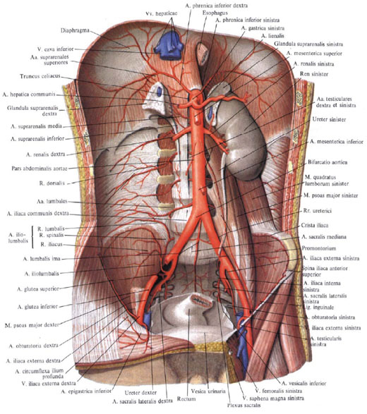
1. The ilio-lumbar artery, a. Iliolumbalis, originates from the posterior trunk a. The liaas interna, goes up and back, passes under the large lumbar muscle and at its inner edge divides into the lumbar and iliac branches:
A) lumbar branch, r. Lumbalis, corresponds to the dorsal branch of the lumbar arteries. It is directed posteriorly, gives to the spinal cord a spinal branch, r. Spinalis; Blood supply to the large and small lumbar muscles, the square muscle of the waist, the posterior sections of the transverse abdominal muscle;
B) iliac branch, r. Iliacus, is divided into two branches - superficial and deep.
The superficial branch goes along the iliac crest and, anastomosing with a. Circumflexa ilium profunda, forms an arc from which branches branching, blood supplying the iliac muscle and the lower parts of the muscles of the anterior abdominal wall.
A deep branch gives the branches to the ilium, anastomosing with a. Obturatoria.
2. The lateral sacral artery, a. Sacralis lateralis, going to the medial side, descends the front surface of the sacrum to the inside of the pelvic openings, while it gives away the medial and lateral branches.
Medial branches, only 5-6, anastomose with the branches of the median sacral artery, forming a network.
The lateral branches penetrate through the sacral sacral orifices into the sacral canal, they give here the spinal branches, rr. Spinales, and, going through the dorsal sacral orifices, blood supply the sacrum, the skin of the sacral region and the lower parts of the deep muscles of the back, as well as the sacroiliac joint, pear-shaped, coccygeal muscles and muscle lifting the anus.
3. Upper gluteal artery, a. Glutea superior, is the most powerful branch of the internal iliac artery. As a continuation of the posterior trunk, it emerges from the pelvic cavity through the peri-conical aperture back into the gluteal region, gives along the path branches to the pear-shaped, internal locking muscles and the muscle lifting the anus. Coming out of the cavity of the pelvis, the artery divides into two branches - superficial and deep:
A) surface branch, r. Superficialis, is located between the large and middle gluteus muscles and blood supply them;
B) deep branch, r. Profundus, is divided into the upper and lower branches, rr. Superior et inferior. Closing between the middle and the small gluteus muscles, it supplies them and the muscle that stretches the wide fascia, giving a number of branches to the hip joint, anastomosing with a. Glutea inferior and a. Circumflexa femoris lateralis.
4. The lower gluteal artery, a. Glutea inferior, in the form of a fairly large branch, extends from the anterior trunk of the internal iliac artery, descends the anterior surface of the pear-shaped muscle and sacral plexus and exits the pelvic cavity through the podrusia-shaped aperture along with the internal genital artery.
The lower gluteal artery supplies blood to the gluteus maximus, sends the artery accompanying the sciatic nerve, a. Comitans n. Ischiadici, and gives a series of branches to the hip joint and gluteal region skin, anastomosing with a. Circumflexa femoris medialis, posterior branch of the constriction artery, a. Abturatoria, and with a. Glutea superior.
5. Obstructive artery, a. Obturatoria, departs from the anterior trunk of the internal iliac artery, goes along the lateral surface of the small pelvis, parallel to the arcuate line, forward to the occlusion aperture and leaves the pelvic cavity through the occlusion channel.
Variants are described when a. Obturatoria departs from a. Epigastrica inferior or from a. Iliaca externa.
Before entering the obstructive canal, the occlusion artery gives the pubic branch, and in the canal itself it divides into its terminal branches - the anterior and posterior branches:
A) pubic branch, r. Pubicus, rises on the posterior surface of the upper branch of the pubic bone and, reaching pubic adhesion, anastomizes with the pubic branch of the lower epigastric artery;
B) anterior branch, r. Anterior, goes down the external occlusive muscle, blood supply to it and upper sections of the adductor muscles of the thigh;
C) posterior branch, r. Posterior, is guided posteriorly and downwards along the outer surface of the occlusion membrane and blood supply to the external and internal blocking muscles, the ischium and sends the acetabular branch to the hip joint, r. Acetabularis. The latter, through a notch of the acetabulum, enters the cavity of the hip joint and along the ligament of the femoral head reaches the head of the femur.

Comments
When commenting on, remember that the content and tone of your message can hurt the feelings of real people, show respect and tolerance to your interlocutors even if you do not share their opinion, your behavior in the conditions of freedom of expression and anonymity provided by the Internet, changes Not only virtual, but also the real world. All comments are hidden from the index, spam is controlled.