Autophagy

Autophagy (from other Greek-auto-self and "is") is a process in which internal components of a cell are delivered inside its lysosomes (in mammals) or vacuoles (yeast cells) and are degraded in them.
The very word "autophagy" means "eating yourself". This is an integrated mechanism for waste disposal - unnecessary proteins, old, worn out cells and other waste biomaterial, as well as bacteria and viruses destroyed by the immune system. Without this process, our cells would have died under an avalanche of dead bacteria and viruses that had worked out their proteins and worn out organelles.
Types and mechanisms of autophagy

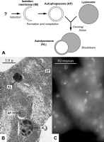
A: Autophagosome formation pattern: an isolating membrane surrounds the cell structures and creates an autophagosome (AP) that fuses with the lysosome and creates autolysosome (AL).
Q: Electron microphotography of autophagosome structures in the fat body of the Drosophila larva. C: Marked by the fluorescent label of the autophagosome in the cells of the liver of a fasting mouse
Now three types of autophagy are distinguished: microautophagy, macroautophagy and chaperone-dependent autophagy. In microautophagy, macromolecules and fragments of cell membranes are simply captured by the lysosome. In this way, the cell can digest proteins when there is a lack of energy or building material (for example, when fasting). But the processes of microautophagy occur under normal conditions and are generally non-selective. Sometimes organoids are digested during microautophagy; Thus, in yeast, microautophagy with peroxisomes and partial microautophagy of nuclei are described, in which the cell remains viable.
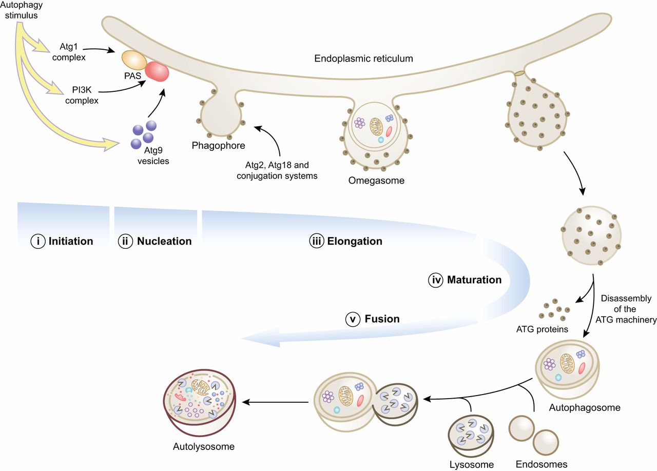
In macroautophagy, the cytoplasmic site (often containing some organoids) is surrounded by a membrane compartment, similar to a cistern of the endoplasmic reticulum. As a result, this region is separated from the rest of the cytoplasm by two membranes. Such two-membrane organelles surrounding the removed organelles and the cytoplasm are called autophagosomes. Autophagosomes combine with lysosomes, forming autophagolysomes, in which the organelles and the rest of the contents of autophagosomes are digested.
Apparently, macroautophagy is also indiscriminate, although it is often emphasized that with the help of this cell the cell can get rid of the "organellids that have served their time" (mitochondria, ribosomes, etc.).
The third type of autophagy is chaperone-mediated. With this method, there is a directional transport of partially denatured proteins from the cytoplasm through the lysosome membrane to its cavity, where they are digested. This type of autophagy, described only for mammals, is induced by stress. It occurs with the participation of cytoplasmic chaperone proteins of the hsc-70 family, accessory proteins and LAMP-2, which serves as the membrane receptor of the chaperone complex and the protein to be transported to the lysosome.
In the autophagic type of cell death, all cell organelles are digested, leaving only cellular debris absorbed by macrophages.
Regulation of autophagy
Autophagy accompanies the vital activity of any normal cell under normal conditions. The main incentives for strengthening the processes of autophagy in cells can be
- Lack of nutrients
- Presence of damaged organelles in the cytoplasm
- Presence in the cytoplasm of partially denatured proteins and their aggregates
In addition to starvation, autophagy can be induced by oxidative or toxic stress.
At present, genetic mechanisms regulating autophagy are studied in detail by yeast. Thus, the formation of autophagosomes requires the activity of numerous proteins of the Atg family (autophagosome-related proteins). Homologues of these proteins are found in mammals (including humans) and plants.
The importance of autophagy in normal and pathological processes
Autophagy is one of the ways to get rid of cells from unnecessary organelles, as well as the body from unnecessary cells.
Especially important is autophagy in the process of embryogenesis, with the so-called self-programmed cell death. Now this variant of autophagy is often called caspase-independent apoptosis. If these processes are violated, and the destroyed cells are not removed, then the embryo most often becomes unviable
Sometimes, thanks to autophagy, the cell can fill the lack of nutrients and energy and return to normal life. On the contrary, in the case of intensification of autophagy processes, cells are destroyed, and their place is in many cases occupied by connective tissue. Such violations are one of the causes of heart failure.
Violations in the process of autophagy can lead to inflammatory processes if parts of dead cells are not removed.
Especially large (though not fully understood) role of autophagy disorders play in the development of myopathies and neurodegenerative diseases. So, with Alzheimer's disease in the processes of the neurons of affected areas of the brain, an accumulation of immature autophagosomes is observed, which are not transported to the body of the cell and do not merge with lysosomes. Mutant hantingtin and alpha-sinuclein are proteins, the accumulation of which in neurons causes, respectively, Huntington's disease and Parkinson's disease-are absorbed and digested in chaperone-dependent autophagy, and activation of this process prevents the formation of their aggregates in neurons.
Autophagy: for which they gave the Nobel Prize in Medicine

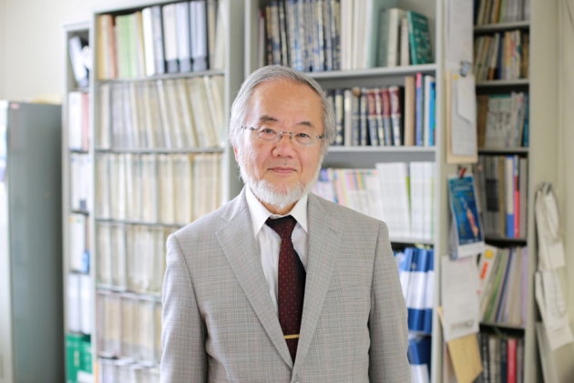
This process was first described in 1963 by the Belgian biologist Christian de Duve, who observed how cells "digest" unnecessary substances with the help of special organelles, which de Duve called lysosomes. However, the mechanisms of autophagy became clear only after the experiments of Yoshinori Osumi (currently he works at the Tokyo Institute of Technology in Yokohama). To determine which genes are responsible for the course of autophagy, Osumah experimented with strains of yeast, in which this process proceeded with a deviation from the norm.
This process was first described in 1963 by the Belgian biologist Christian de Duve, who observed how cells "digest" unnecessary substances with the help of special organelles, which de Duve called lysosomes. However, the mechanisms of autophagy became clear only after the experiments of Yoshinori Osumi (currently he works at the Tokyo Institute of Technology in Yokohama). To determine which genes are responsible for the course of autophagy, Osumah experimented with strains of yeast, in which this process proceeded with a deviation from the norm.
The cell has two ways to get rid of biological "garbage". First, it can take advantage of the huge molecules of ubiquitin-protease, an enzyme that can break down proteins into individual amino acids. Proteases float freely in the cytoplasm. Osumah investigated the second method - splitting "garbage" in lysosomes; It is called autophagy.

The process of autophagy: first around the object - molecules, parts of the organelle, organelles, bacteria or virus, a membrane is formed. When it closes, the formed autophagosome transports the object to the lysosome and combines with it into the autolysosome, where the splitting of the object takes place.
Autophagy is realized as follows: first, the cell sequester (isolates) the molecules to be destroyed, lining the membrane around them. The resultant quasi-organelle with a molecule or even another organelle enclosed in it is called an autophagosome. The autophagosome then converges with the lysosome, forming an autolysosome. Inside it, an isolated object is exposed to hydrolases, enzymes that dissolve it.
Autophagy is a normal part of the cell's life cycle. It ensures cell survival during starvation, makes cell differentiation and growth control possible, and also participates in the regulation of many other processes in the cell. Incorrect leakage of autophagy is associated with the development of Parkinson's disease, type 2 diabetes and other diseases. However, the practical application of the discovery of Osumi and his colleagues have not yet been found.
Via wikipedia.org & popmech.ru


Comments
When commenting on, remember that the content and tone of your message can hurt the feelings of real people, show respect and tolerance to your interlocutors even if you do not share their opinion, your behavior in the conditions of freedom of expression and anonymity provided by the Internet, changes Not only virtual, but also the real world. All comments are hidden from the index, spam is controlled.