Parameters and transcript of the cardiac eq

Electrocardiography is a technique for recording and examining the electric fields produced by the heart. Electrocardiography is a relatively inexpensive, but valuable method of electrophysiological instrumental diagnostics in cardiology. The direct result of electrocardiography is the production of an electrocardiogram (ECG).
To interpret the electrocardiogram means to evaluate the activity of the cardiac muscle biopotential. This allows the doctor to detect a violation of the rhythm frequency, ischemia, ventricular hypertrophy, atrial and a number of other anomalies.
The process of decoding the ECG (data of the cardiographic pattern) consists in measuring the length, the magnitude of the segments, and the amplitude of the oscillation of the teeth.
A study of the results of a healthy person will help to compare the data, and to identify the existing problems of the heart in pathological changes.

Electrocardiography (ECG) is a non-invasive test, which provides valuable information on the condition of the heart.
The essence of this method consists in recording the electrical potentials that arise during the operation of the heart and in their graphic display on a display or paper.
Decoding ECG of the heart
To begin with, consider the decryption plan, for this we need to establish:
- The nature of the heart rate and the determination of the exact value of the contractions in the time interval
- Cycle of cardiac biopotentials
- Recognition of excitation sources
- Conductivity evaluation
- The study of the P wave and the QRST ventricular interval
- The designation of the axis of propagation of signals and the position of the heart relative to its
The work of the heart is determined by the emerging biopotentials.
ECG decoding is a graphical representation of the intensity of a given discharge, which helps to identify malfunctions in the operation of the cardiac divisions.
The rhythm of the contractions of the heart muscle is determined by the duration of the measurement of the RR intervals. If their duration is the same or is marked by fluctuations of 10% - this is considered the norm, in other cases, one can speak of a rhythm disturbance.
ECG indicators and their interpretation

Heart rate (heart rate)
Let's list the basic parameters of ECG, which interest us on the cardiogram:
- Zubtsy - characterize the stages of the cardiac cycle
- 6 leads - heart departments displayed in numbers and letters
- 6 thoracic - fix changes in cardiac potentials in the horizontal plane

The PQ QRS QT interval displays the impulse conductivity
After reading the terminology, you can try to decipher the results on your own. However, we remind that 100% objective diagnosis can be made only by the attending physician.
The height of the teeth begins to measure from the contour line - the horizontal line using a ruler, taking into account the location of the positive teeth above the straight line and the negative ones below the axis.
Their shape and dimensions depend on the passage of the electric wave and differ in all leads. By automatically specified formula we calculate the duration of intervals and segments - divide the distance between segments by the speed of the tape.
Values of teeth on the cardiogram
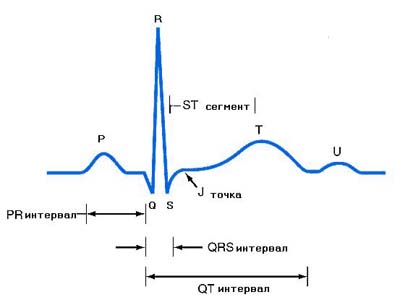
The tooth P - is responsible for the propagation of the electrical signal in the atria. Norm: positive value with a height of up to 2.5 mm.
For the Q wave, the impulse distribution along the interventricular septum is characteristic. Norm: always negative, and often not recorded by the device because of its small size. Its severity is cause for concern.
The tooth R is considered to be the largest. It reflects the activity of the electric pulse in the myocardium of the ventricles. His wrong behavior indicates hypertrophy of the myocardium. The interval norm is -0.03 s.
Sine S - shows the completeness of the excitation process in the ventricles. Norm: negative and does not exceed 20 mm.
Interval PR - indicates the rate of distribution of excitation at the atrium to the ventricles. Norm: the oscillation is 0.12-0.2s. This interval determines the heartbeat.
Tine T - reflects the repolarization (recovery) of the biopotential in the cardiac muscle. Norm: positive, duration - 0,16-0.24 s. Indications are informative for diagnosing ischemic abnormalities.
Interval TR - shows a pause between abbreviations. Duration - 0.4 sec.
Segment ST - differs by maximum excitation of the ventricles. Norm: allow a deviation of 0.5 -1mm down or up.
QRST Interval - Displays the time period for the excitation of the ventricles: from the beginning of the passage of the electrical signal and until their final contraction.
Decoding ECG in children
The norms of children's testimony are significantly different from those of adults. For ECG transcripts in children, the curve should be tracked and the numerical parameters of the teeth and intervals should be compared.
The norm is:
- Deep position of the tooth Q
- Sinus arrhythmia
- The QRST ventricular interval is subject to an alternative (reversal of the T-wave polarity)
- Atrial movement of the source of rhythm is noted
- With the child's growing, the number of thoracic leads with a negative tooth T decreases
- The large dimensions of the atria determine the height of the tooth P
- The age of the child affects the intervals of the ECG - they become longer. Small children are dominated by the right ventricle
Sometimes the intense growth of the baby provokes violations in the cardiac muscle, which can show a cardiogram.
What does sinus rhythm on a cardiogram mean?
Decoding the ECG shows a sinus rhythm? This indicates the absence of pathologies, and is considered the norm with a characteristic stroke rate from 60 to 80 per min. With an interval of 0.22 s. The presence of a doctor's record about the irregularity of the sinus rhythm involves fluctuations in pressure, dizziness, and chest pain.
Rhythm, indicated by 110 strokes, indicates the presence of sinus tachycardia. The cause of its occurrence may be physical exertion or nervous excitability. Such a condition can be temporary, and does not involve long-term treatment.
With anemia, myocardium, or fever, there is a persistent manifestation of tachycardia with rapid heartbeat. Decoding the ECG in this case determines the unstable sinus rhythm, and indicates an arrhythmia - an increased frequency of contractions of the cardiac divisions.
For children, too, this symptom is characteristic, but the sources of origin are different. These are cardiomyopathy, endocarditis and psychophysical overloads.
The rhythm can be disturbed from birth, have no symptoms, and come to light during electrocardiography.
Via vse-o-gipertonii.ru


Comments
When commenting on, remember that the content and tone of your message can hurt the feelings of real people, show respect and tolerance to your interlocutors even if you do not share their opinion, your behavior in the conditions of freedom of expression and anonymity provided by the Internet, changes Not only virtual, but also the real world. All comments are hidden from the index, spam is controlled.