Decoding of heart electrocardiography: ECG norm

Electrocardiography is a technique for recording and examining the electric fields produced by the heart. Electrocardiography is a relatively inexpensive, but valuable method of electrophysiological instrumental diagnostics in cardiology. The direct result of electrocardiography is the production of an electrocardiogram (ECG).
We continue our series of articles about the ECG. In this article, we consider, in the opinion of the editorial board, the most important topic - the norms of electrocardiography indicators. We will review the standards for adults and children, and also try to understand the reason for the discrepancy between the indicators. Article replenished - stay tuned.
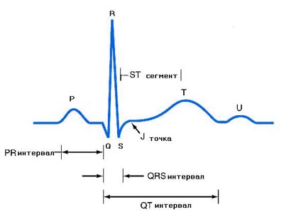
The heart readings are recorded in the electrocardiogram, expressed in the form of teeth and horizontally arranged segments and intervals. The teeth are on an isoelectric line, they are drawn up and down and resemble a curve.
They are marked with Latin letters P, T, S, Q, R, and are written down by the line of the horizontal segment between the teeth T and P at rest.
When decoding the cardiac ECG, the norm is between TQ or TP. It determines the amplitude of oscillations in the duration of the teeth, intervals and width.

Electrocardiography (ECG) is a non-invasive test, which provides valuable information on the condition of the heart.
The essence of this method consists in recording the electrical potentials that arise during the operation of the heart and in their graphic display on a display or paper.
Parameters of normal cardiogram
When interpreting the result, it is necessary to follow the sequence. First pay attention to:
- Rhythm of the heart muscle
- Conductivity of intervals
- Electric axis
- Analysis of QRS complexes
- ST segments and tooth T
Decoding of the cardiac ECG for the determination of the norm is reduced to the indications of the position of the teeth. The normal heart rate is measured by the length of the RR intervals (the distance between the high denticles). They should be the same and do not exceed the difference of 10%.
The rapid rhythm indicates a tachycardia, a slowed-down bradycardia. The norm is 60-80 pulses per minute.
On the intervals of P-QRS-T, located between the teeth, one can judge the pulse passing through the cardiac divisions. The interval norm is 120-200 ms or 3-5 squares.
Interval PQ will show the penetration of the bio-potential to the ventricles through the ventricular node directly to the atrium.
The QRS complex will give an idea of the excitation of the ventricles. To do this, measure the width of the complex from the tooth Q to the S-tooth. Norm - the width is 60 -100 ms.
Decoding of the cardiac ECG is the norm of the tooth Q, which does not exceed the value in duration - 0, 04 and 3 mm in depth.
The QT interval informs about the duration of ventricular contraction. The norm is 390 -450 ms. A longer interval indicates ischemia, atherosclerosis or myocarditis and rheumatism, shortened - about hypercalcemia.
When decoding the ECG, the normal electric axis of the heart reveals the impulse conduction disturbance zones. Values are calculated automatically.
To do this, you need to monitor the height of the teeth. Norm: the tooth S should not be higher than the R wave. If there is a deviation to the right, in the withdrawal, the height of the S-tooth is smaller than the R-tooth, this indicates a deviation in the right ventricle. Such a deviation to the left is a tooth S above the R-tooth, which determines left ventricular hypertrophy. The QRS complex shows the passage of the biopotential through the septum and the myocardium. Norm: no tooth Q or more than 20-40ms in width and 1/3 of the tooth R.
The segment ST is measured from the end of the tooth S until the T-tooth begins. The pulse rate affects the duration of the ST. Segment rate: permissible deviations from the isoline - 0,5 mm in depression, lifting in the leads - up to 1 mm.
Read the battlements
Normally the tooth P is positive in the I and II leads, in VR - negative. Width up to 120ms. Reflects the picture of the distribution of the biopotential in the atria. Negative T in I and II are signs of ischemia, ventricular hypertrophy, infarction.
The tooth Q-fixes the excitation of the left half of the septum. Norm: 1/4 of the R wave at 0.3 s. An increase in the norm indicates a necrotic pathology of the myocardium.
The tooth R - reflects the activity of the walls of the ventricles of the heart. Normally recorded in all leads. In the presence of another picture, one can judge the hypertrophy of the ventricles.
Sine S - shows excitation of septa and basal ventricular layers. The height of the tooth is 20 mm. Attention is drawn to the ST segment, which determines the condition of the heart. Fluctuations in the position of the segment indicate ischemia of the myocardium.
The tine T is marked upward in the leads I and II, in the VR leads it is constantly negative. Acute high T - the index of hyperkalemia, flat and long - hypokalemia.
ECG transcript in children: norm
- The frequency of heart attacks in children (heart rate) to 3 years is 100 -110 pulsations
- From 3 to 5 years - 100 strokes, in adolescents - from 60 to 90
- The norm of the tooth P is not higher than 0, 1c
- The QRS complex is indicated by readings from 0.6 to 0.1 s
- Intervals PQ -varying from 0.2 to 0.2, QT to 0.4 s
- Electric axis unchanged
- Rhythm of sinus
ECG indices when interpreting the sinus rhythm rate expresses the dependence of the pulse rate on respiration. This means that the rhythm of the heart muscle is normal, and is 60 -80 beats per minute. In the interval, the QRS has a prong P of the correct shape.
Causes of different ECG indices
The patient's ECG readings may sometimes differ. To obtain accurate results, many factors need to be taken into account.
Often the distortion of results is due to technical defects. This is possible if the resulting cardiogram is not correctly glued together. Confusion occurs because of Roman numerals, which are the same in the correct and inverted values.
Quite often problems arise when cutting a graph, where the first tooth P or the last T
Significance has a preliminary preparation for the procedure (read how to prepare for the ECG).
The impact of electrical appliances working in the neighboring rooms affects the oscillations of the alternating current, which is reflected by the repetition of the teeth.
Excitement of the patient or uncomfortable position during the session affects the instability of the zero line.
The possibility of an inadvertent arrangement of the electrodes or their displacement is not ruled out.
Measurements on the multichannel electrocardiograph are the most accurate.
Via vse-o-gipertonii.ru


Comments
When commenting on, remember that the content and tone of your message can hurt the feelings of real people, show respect and tolerance to your interlocutors even if you do not share their opinion, your behavior in the conditions of freedom of expression and anonymity provided by the Internet, changes Not only virtual, but also the real world. All comments are hidden from the index, spam is controlled.