
| Start of section
Production, amateur Radio amateurs Aircraft model, rocket-model Useful, entertaining |
Stealth Master
Electronics Physics Technologies Inventions |
Secrets of the cosmos
Secrets of the Earth Secrets of the Ocean Tricks Map of section |
  |
| Use of the site materials is allowed subject to the link (for websites - hyperlinks) | |||
Navigation: => |
Home / Patent catalog / Catalog section / Back / |
|
INVENTION
Patent of the Russian Federation RU2164687
![]()
METHOD FOR DIAGNOSTICS OF EXTERNAL GENITAL ENDOMETRIOSIS
The name of the inventor: Posiseeva LV; Sotnikova N.Yu .; Shor AL; Antsiferova Yu.S.
The name of the patent holder: Ivanovo Research Institute of Maternity and Childhood. V.N. Gorodkov
Address for correspondence: 153731, Ivanovo, ul. Victory 20, Research Institute of the Ministry of Health of the Ministry of Health
The date of the beginning of the patent: 1999.05.17
The invention relates to medicine, in particular to gynecology. The method provides an increase in the sensitivity and specificity of the study, as well as the accuracy of the diagnosis. The method is simple and generally available. The peripheral blood of the patient is examined. Spontaneous HCT activity of neutrophils after incubation with PAMG-2 is determined and at 24% and beyond, external genital endometriosis is diagnosed.
DESCRIPTION OF THE INVENTION
The invention relates to medicine, namely gynecology, and can be used for the diagnosis of external endometriosis.
The urgency of the invention is determined by a high (up to 17%) frequency of occurrence of external endometriosis [1, 2]. External endometriosis is found in 40 - 48% of infertile women [2]. External endometriosis increases the risk of infertility by 20 times [10]. The diagnosis of external genital endometriosis, as a rule, is established on the basis of the results of invasive instrumental studies (laparoscopy or laparotomy with subsequent histological examination of the removed tissues). In this regard, the development of reliable laboratory diagnostic criteria for endometriosis presents an urgent task for practical public health.
A method for diagnosing external endometriosis is known based on the determination of the content of autoantibodies in the serum of peripheral venous blood to endometrial cells, the essence of which is that an increase in the titer of serum autoantibodies is established and a severe endometriosis of grade III and IV is diagnosed with an accuracy of 45.5% [9].
Disadvantages of the method:
- low accuracy (45.5%),
- this method allows to diagnose only severe stages of endometriosis, which occur in less than 30% of cases, diagnosis of which is possible on the basis of primary examination, history and ultrasound results,
- The method is not informative at stages I and II of the disease.
The method closest to the claimed method is the method for diagnosing external genital endometriosis by examining a biological material, characterized in that basophilic leukocytes are isolated from the patient's blood, spontaneous release of histamine is determined and at this 49% or more, external genital endometriosis is diagnosed [5] .
This method was chosen by us as a prototype. However, it has a number of drawbacks:
- the release of basophilic leukocytes requires the collection of large amounts of blood (18 ml) because of the relatively small number of basophils in human peripheral blood (0.5-1%) and a low content of histamine in them [4];
- the proposed method is non-specific, since an increase in histamine release by basophils occurs in other pathological conditions, for example, in allergic reactions of immediate type;
- there is no data on the accuracy of the method.
These disadvantages are proposed to be eliminated in the claimed method.
The claimed technical result is achieved by the fact that in the peripheral blood the index of spontaneous HCT activity of neutrophils is determined after their incubation with placental a -2 microglobulin of fertility (PAMG-2) and at the values of this index equal to 24% and more, external genital endometriosis is diagnosed with accuracy 84%.
The indicator of HCT activity of peripheral neutrophils is based on the ability of phagocytes to utilize oxygen with formation of free radicals and serves as an indicator of a respiratory "explosion" in cells. Previously, it was used as one of the diagnostic criteria of the inflammatory process, to control the effectiveness of antibiotic therapy [6]. It is known that with external endometriosis the content of PAMG-2 in serum increases [8]. This probably serves as a factor sensitizing the neutrophils of peripheral blood of women with endometriosis.
The novelty of the claimed method is that for the first time it is proposed to diagnose external endometriosis by determining the parameters of the HCT activity of peripheral neutrophils after their incubation with PAMG-2.
The method is carried out as follows:
By a standard procedure, neutrophils from 1 ml of heparinized blood from the ulnar vein are isolated [7]. Next, we propose to add to the 0.1 ml suspension of cells at a concentration of 10 6 cells / ml an equal volume of Medium 199 containing 20 ng of PAMG-2. The cells are incubated for 60 min at t = 37 ° C in a thermostat, washed with Medium 199 by centrifugation for 10 minutes and put a spontaneous HCT test according to a standard procedure [3]. The parameters of the neutrophil NST activity are calculated by light microscopy.
Using the proposed method allows to diagnose external genital endometriosis with an accuracy of 84%.
Distinctive features of the method are:
- the diagnostic parameter of the HCT activity of peripheral blood neutrophils, determined after the incubation of neutrophils with PAMG-2, with a quantitative value of 24% or more, is diagnosed with external genital endometriosis.
The essence of the claimed method is explained by the following examples:
Example 1. Patient P., 25 years old. Turned about the secondary infertility of the expressed pain syndrome in the form of algodismenorea, dyspareunia. It is sick about 6 years. In the history of medical abortion. Repeatedly carried out anti-inflammatory therapy in occasion of exacerbation of chronic 2-sided adnexitis without effect. Objectively: the uterus of normal size, deflected posteriorly, the attempt to bring it to its normal position is sharply painful, a painful dense knot 1.5 cm in diameter is palpable in the rectovaginal septum. Attachments without features. Palpation of the sacro-uterine ligaments is painful. At a finger examination of the rectum, no pathology was detected.
Clinical diagnosis: external and retrocervical endometriosis.
According to the immunological examination according to the claimed method, the indicator of spontaneous HCT activity of neutrophils after incubation with PAMG-2 was 38%.
Conclusion: according to the claimed method, external endometriosis is diagnosed.
On the 7th day of the menstrual cycle, laparoscopy was performed, in which deep foci of endometriosis, an area of more than 3 cm 2 on the peritoneum of the sacro-ovaries and an infiltrating focus in the rectovaginal septum with cicatricial changes, were detected. Excision of endometriosis foci with a monopolar coagulator and neurotomy was performed. The histological examination of the removed tissues confirmed the diagnosis.
Example 2. Patient L., 36 years old. Turned about secondary infertility. In the history: 1 timely delivery, 2 medical abortions without complications, IUD for 4 years. At present he is in his second marriage, he does not use contraception for three years, pregnancy does not occur. At the time of the examination, he complains of moderate pulling pains in the lower abdomen, which intensify during menstruation. Examined for infertility: the cycle is biphasic, according to hysterosalpingography, the uterus is not changed, the uterine tubes are moderately convoluted, freely passable. The husband was examined and healthy. Miller's test is positive.
With vaginal examination the body of the uterus is slightly more than normal size, in the lateral arches painful tightness.
Clinical diagnosis: chronic salpingitis, external endometriosis. A survey using the claimed method showed that the spontaneous HCT activity of neutrophils incubated with PAMG-2 was 12%.
Conclusion: according to the claimed method, the diagnosis of endometriosis is not confirmed.
On laparoscopy, the uterus and uterine tubes are not changed, the pelvic peritoneum without pathological inclusions. In the right ovary is the preovulatory follicle. As a cause of pain syndrome, varicose veins of parametres on both sides are revealed. The conducted intraoperative postcoital test revealed the cause of infertility: no aspirated peritoneal fluid and posterior third of the cervical canal of mobile spermatozoa were found.
Example 3. Patient S., 22 years old. Appealed about the primary infertility. It is in the first marriage, there were no gynecological diseases in the anamnesis, there are no specific complaints.
In the pre-operative examination, a two-phase menstrual cycle is established, the "male" factor is excluded, the postcoital test is positive. According to hysterosalpingography, the uterine cavity has the right shape, the left fallopian tube is not contrasted, the right uterine tube is filled up to the ampullar region, there is no contrast in the free abdominal cavity.
With an objective examination of the uterus of normal size, position. Appendages are not determined, the vaults are free, mucous membranes, uninvolved.
Conclusion: chronic salpingitis with occlusion of the fallopian tubes.
According to the survey data on the claimed method, the spontaneous HCT activity of neutrophils after incubation with PAMG-2 was 24%.
Conclusion: according to the claimed method, external genital endometriosis is suspected.
With laparoscopy, the uterine tubes are not changed, and with chromohydrotubation they are freely passable. On the peritoneum of the posterior leaf of the wide uterine ligament on the left and the surface of the left ovary, single foci of endometriosis were found. Foci on the peritoneum are coagulated by a bipolar coagulant, the ovary is resected. The result of the histological examination of the drug is the ovarian endometriosis, which confirms the correctness of the diagnosis, established according to the claimed method.
In this way, 19 patients were examined, the data are given in Table 1.
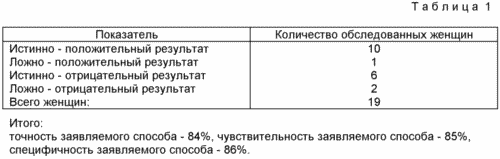
Advantages of the claimed method:
1. High accuracy - 84%, sensitivity - 85%, specificity - 86%.
2. A small amount (1 ml) of blood needed for the study.
3. Simplicity, accessibility of the methodology.
BIBLIOGRAPHY
1. Baskakov V.P. Clinic and treatment of endometriosis. - M., 1990.- 240 p.
2. Genital endometriosis: clinic, diagnosis, treatment (methodical recommendations). - Moscow, 1997. - 31 p.
3. Demin A.A. NTB - test for bacterial and non-bacterial diseases // Sov. Medicine. - 1976. - N 12. - P. 16-20.
4. Immunological methods / Ed. G. Fremelya .- M .: Medicine, 1987. - P. 358-361.
5. Ionov ID, Volkov NI, Sukhikh GT, Pshenichnikova T.Ya. The method of diagnosis of external genital endometriosis // Discoveries of the invention. - 1990, No. 1. - P. 180 (AS No. 1534397).
6. Mayanskii AN, Pikuza O.I. Clinical aspects of phagocytosis. - Kazan: Magarif, 1993. - 192 p.
7. Petrova IV, Vasilyeva LA A method for isolating from a human peripheral blood a pure population of neutrophils to study their rosette-forming properties // LAB. - 1974. - N 11. - P. 26-28.
8. Tatarinov Yu. S., Posiseeva L.V., Petrunin D.D. Specific alpha 2- microglobulin (glycodelins) of the human reproductive system: 20 years from basic research to introduction into clinical practice. - Ivanovo, MIK, 1998. - 128 p.
9. Martinez P., Ulcova-Gallova Z., Iborra A., Palacio JR, Bousel L., Rokyta Z., Novotny Z. Endometriosis and autoantibodies // Am. J. Reprod. Immunol. - 1998. - v.40. - N 4. - p. 271.
10. Ory SJ Pelvic endometriosis // Obstet. Ginnecol. Clin. J. Amer. - 1987. - Vol. 14, No. 4. - P. 999-1014.
CLAIM
A method for diagnosing external genital endometriosis by examining peripheral blood, characterized in that the spontaneous HCT activity of neutrophils is determined in the blood after incubation with PAMG-2 and at the rates of 24% and more, external genital endometriosis is diagnosed.
print version
Date of publication 27.03.2007gg
![]()

Comments
When commenting on, remember that the content and tone of your message can hurt the feelings of real people, show respect and tolerance to your interlocutors even if you do not share their opinion, your behavior in the conditions of freedom of expression and anonymity provided by the Internet, changes Not only virtual, but also the real world. All comments are hidden from the index, spam is controlled.