
|
Start of section
Production, amateur Radio amateurs Aircraft model, rocket-model Useful, entertaining |
Stealth Master
Electronics Physics Technologies Inventions |
Secrets of the cosmos
Secrets of the Earth Secrets of the Ocean Tricks Map of section |
  |
| Use of the site materials is allowed subject to the link (for websites - hyperlinks) | |||
Navigation: => |
Home / Patent catalog / Catalog section / Back / |
|
INVENTION
Patent of the Russian Federation RU2196497
![]()
DEVICE FOR DIAGNOSTICS OF THE PATHOLOGY OF THE VISUAL SYSTEM IN CHILDREN AT THE CRITICAL FREQUENCY OF MILKANIUM FUSIONING
The name of the inventor: Golubtsov K.V . ; Sofronov PD
The name of the patent holder: Institute for Information Transmission Problems of the Russian Academy of Sciences; Golubtsov Konstantin Vasilievich
Address for correspondence: 129010, Moscow, ul.B. Spasskaya, 25, building 3, LLC "Law Firm Gorodissky and Partners", Yu.D. Kuznetsov, registration number 595
Date of commencement of the patent: 2000.11.14
The invention relates to the field of medical technology, namely, to devices for diagnosing the visual system. The device comprises a housing in which a means for generating pulses is arranged, the outputs of which are electrically connected to two light stimulators made in the form of light-emitting diodes fixed in a light shield which is installed in the headlights of a toy car and a power unit that is electrically connected to the means for generating pulses. The means for generating pulses is made in the form of a microprocessor, by means of which pulses of an adjustable frequency are generated in the range from 1 to 60 Hz. The device contains both an IR transceiver installed in the housing, connected to a microprocessor for receiving and transmitting data, and an indicator connected to the microprocessor for indicating the indication of the critical flicker frequency for the right and left eyes of the child. On the control panel there are control buttons. The invention makes it possible to simplify the diagnostic procedure.
DESCRIPTION OF THE INVENTION
The invention relates to ophthalmology, and more specifically to a device for diagnosing the pathology of the visual system in children at a critical flicker frequency.
The invention can be used to diagnose the visual system in children, and as an integral part of the device, children's toys can be used, in particular a toy car, in which a diagnostic tool is built into the headlights. The invention can be used for differential diagnosis of the functional state of the retina and optic nerve, the pathology of the optic nerve.
It is known that an image that is immovable relative to the retina a person ceases to see after 1-3 seconds, and visual perception occurs only under conditions of a change in brightness, chromaticity or the position of the object. Information on stationary objects or during prolonged exposure is transmitted along the visual pathway only due to eye tremor, which occurs at a frequency of 30 Hz. Inadequate long-term light and color effects-exposure and color music-cause certain disorders of the retina and optic nerve to disrupt photochemical and metabolic processes in the retina.
Light perception and color perception are the basis on which other functions of vision are built. The most important of these is the distinction and recognition of the form of objects. For its implementation, it is not the absolute sensitivity of the eye to light and color that is important, but the sensitivity to their changes in space and time is the so-called contrast sensitivity, expressed by the numerous quantitative relationships established by the experimental path-the psychophysical laws of vision.
There are contrast sensitivity - spatial and temporal. Temporary contrast sensitivity is the minimum frequency of flickering of stimuli per unit of time, at which the phenomenon of fusion of individual visual sensations already occurs. This phenomenon is called the critical frequency of flicker fusion (CFE). Currently, in ophthalmology, the use of time-frequency indicators, which expand information, reveal features of the nature of vision and its functional disorders, is becoming increasingly important.
CSFM does not depend on visual acuity, on refraction and on the size of the pupil. However, it is established that CFCC changes under the influence of side stimuli: sound, olfactory, taste and tactile.
In pediatric ophthalmology very often early diagnosis of the pathology of the retina and optic nerve causes difficulties in connection with the microsymptomatic picture of clinical signs on the fundus, which leads to diagnostic errors. In order to clarify the diagnosis and differential diagnosis, it is necessary to carry out additional psychophysical and electrophysiological studies, which are often difficult for children to carry out, and now are very expensive. Currently, frequency and time methods are preferred. The devices used in this case are compact and easy to use.
A method for diagnosing the degree of man's fatigue is known (see, for example, SU 1445694 Ovchinnikova ND, A 61 V 5/16) , that is placed in front of the patient's eyes a light-protective eyeglass with integrated LEDs in the front panel connected to A means for generating and forming pulses of light of different wavelengths, light pulses of green blue and red are applied to the LEDs, and diagnostics of the functional state of the visual system is performed.
Light flashes are simultaneously applied to both eyes paracentrically and the degree of interhemispheric functional asymmetry of the brain is determined by the difference in the critical frequency of flicker fusion, which is used to judge the degree of fatigue of the patient being examined. However, the proposed method does not allow to perform diagnostics of the functional state of the visual system and, in particular, the central zone of the retina, excludes the use of color stimuli and does not determine their significance. The method does not allow simultaneous restoration of visual functions.
This method is difficult to use to diagnose the pathology of the visual system in a child, because the child is often afraid to wear a sun-protecting eyeglass frames with built-in LEDs. The child also can not give the exact answer to questions of the doctor at diagnostics.
A device is known for diagnosing and restoring visual functions (see, for example, RF patent No. 2071301) containing a housing in which a pulse generator is located whose outputs are connected to two light stimulators fixed in a light shield and a power supply that is electrically connected to a pulse generator .
Using this device, the patient is exposed to light from one wavelength, lying in the visible region of the spectrum of 400-700 nm, with an illumination of 5 to 100 lux. In this case, one or both eyes are exposed to flashing light with a flashing frequency of 0.06 - 1 Hz. This device will contain glasses with light-insulated eyepieces, each of which has a light-diffusing reflector, inside of which a light source and mounted opposite it with the ability to replace the light filter are fixed.
The device contains both a frequency control of the flashing of light, a brightness control for light, a switch providing a given light effect on each eye, and a power supply.
This device is difficult to use to diagnose the pathology of the visual system in a child, because the child is often afraid to wear a sun-protecting eyeglass frame with built-in LEDs. The child also can not give the exact answer to questions of the doctor at diagnostics.
The present invention is based on the task of creating a device for diagnosing the pathology of the visual system in children at a critical flicker frequency in which the arrangement of the device elements in the body and in the headlights of a toy car, the communication between which is carried out by means of infrared radiation, will allow diagnosis of the child's visual system , I.e. Functional state of the retina and optic nerve.
The problem is solved by the fact that in an apparatus for diagnosing the pathology of the visual system in children at a critical flicker frequency containing a housing in which a means for generating pulses is located, the outputs of which are electrically connected to two light stimulators made in the form of LEDs fixed in a light shield, Which is installed in the headlights of a toy car, and the power supply unit which is electrically connected to the means for generating pulses, according to the invention, the means for generating pulses is implemented in the form of a microprocessor by means of which pulses of an adjustable frequency are generated in the range of 1 to 60 Hz, and the device comprises a transceiver On the IR-radiation, installed in the housing, connected to the microprocessor and intended for receiving and transmitting data, connected to the microprocessor indicator for indicating the indication of the critical flicker frequency for the right and left eyes of the child, connected to the microprocessor control panel with the switch on which the button Control for switching on the microprocessor, the button for adjusting the luminance of the light stimulus, the button for switching the color of the LEDs, the button for activating the transceiver on the infrared radiation, for switching the indications on the indicator for the left and right eyes, the button for connecting one of the light stimulators to the power supply unit installed in the car; The housing of the toy car is connected in series with a second IR transceiver connected to a power supply unit for receiving and transmitting data from the microprocessor and a second microprocessor for forming a pulse duration of the adjustable frequency and stabilizing the current applied to the LEDs connected to the unit And electrically connected to the first and second light stimulators.
It is useful that the device contains an adapter for connecting the power supply to the electrical network.
It is advisable that each LED is a multi-color LED.
It is advantageous to use LEDs having at least three primary colors selected from the group consisting of red, green and blue.
In the following, the invention is illustrated by a description of preferred embodiments thereof, with reference to the accompanying drawings, in which:
 |
 |
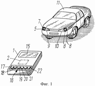
FIG. 1 shows a general view of a device for diagnosing the pathology of the visual system in children at a critical flicker frequency, according to the invention;
FIG. 2 is a block diagram of an apparatus for diagnosing the pathology of the visual system in children at the critical frequency of flicker fusion, according to the invention.
A device for diagnosing the pathology of the visual system in children at the critical flicker frequency comprises a housing 1 (FIG. 1) in which means 2 for generating pulses are located, the outputs 3, 4 of which are electrically connected to two light stimulators 5, 6 made in the form of LEDs, Which are installed in the headlamps 9, 10 of the toy car 11. The device also includes a power supply 12 that is electrically connected to the means 2 for generating pulses.
A power unit 13 is mounted on the vehicle 11.
The means 2 for generating pulses is made in the form of a microprocessor, by means of which pulses of an adjustable frequency are generated in the range 1-60 Hz.
The device also includes an IR transceiver 14 installed in the cabinet 1 connected to the microprocessor 2 for receiving and transmitting data.
The indicator 15 is connected to the microprocessor to indicate the indication of the critical flicker frequency for the right and left eyes of the child.
A control panel 16 with a switch is also connected to the microprocessor, which includes a control button 17 for activating the microprocessor and the IR transceiver, a light stimulus adjustment button 18, a light color switching button 19, a flickering frequency increase button 20, a frequency reduction button 21 Flicker fusion, button 22 for switching the indications on the indicator for the left and right eyes.
In the case of the toy car 11, a second transceiver 23 is connected in series on the infrared radiation connected to the power unit 13 mounted on the car 11 and intended for receiving and transmitting data from the microprocessor, and a second microprocessor 24 for forming a pulse length of the adjustable frequency connected to the The power supply unit 13 and to the first and second light stimulators 5 and 6.
In a preferred embodiment, the device comprises an adapter 25 for connecting the power supply unit 12 or the power supply unit 13 to an electrical network.
Each light stimulator 5, 6 is a multi-color LED.
The device uses LEDs having at least three primary colors, for example red, green and blue.
The device for diagnosing the pathology of the visual system in children according to the critical frequency of flicker fusion is carried out as follows.
Before testing, according to the age of the child, he is introduced to the toy device and the examination method. It is advisable to conduct a trial definition of CFCM binocularly to clarify the child's understanding of the purpose of the survey without recording the testimony.
Examination is carried out for each eye separately. Children with ametropia are advisable to conduct research in the optimal spectacle correction. A child from a distance of 25-30 cm is presented with a car with headlights on. At the same time one of the headlamps burns constantly, and on the other headlight the light flashes with the constantly increasing frequency of flickering, which the doctor smoothly adjusts with the buttons 20, 21. In the case of monocular pathology of the child, it is advisable to begin the examination of CFM from the eye that sees better.
Start the examination from the eye that sees best, from the red, then make a green color, and then blue, then switch the button 19. At the moment when the child ceases to distinguish the flickering, he immediately must give a signal to the doctor, for example, say "stop." Then, the button 22 switches the device to the other eye.
At this moment, the adjustment of CFCM stops and the measurement result is displayed on the screen. This is the first test of CFM (P1).
Then carry out the study in the reverse order, i.e. Begin testing with the maximum frequency of flicking of color-testing, gradually reducing the frequency of the light signal. At the moment when the child indicates the appearance of flashes of color testing, the doctor fixes the result on the screen (P2).
The result of CFCM of this child is determined by calculating the arithmetic mean of these two values
KCSM = (P1 + P2) / 2.
The parameter P1 is normally higher than P2 as a result of the existing visual inertia in the form of a sequential image.
The CSFM was determined for healthy children by examination on these instruments, the results of which are given in Table 1.

Specific features of changes in CFCs were revealed with different vision pathologies.
The definition of CSFM for color stimuli was carried out in children aged 4 to 15 years - 50 healthy children (100 eyes) and 259 children with diseases (518 eyes) with various eye pathologies. As a result of these studies, the indicators of normal values in healthy children for stimuli of red, green and blue on the claimed device were obtained.
Determination of the critical fusion frequency in children with retinal pathology, optic nerve and amblyopia showed high efficacy (up to 83%) with low-symptomatic changes in the fundus.
The results of the studies showed that CFFF depends on the severity of the lesion of the visual system and is practically independent of visual acuity. Examples of diagnosis of vision of various pathologies are given below.
Example 1. Amblyopia
Children with refractive anisometropic and dysbinocular amblyopia according to the ES classification were examined. Avetisova (1964) with a high, medium and low degree. The results are shown in Table 2.

As a result of the CSFM research on color stimuli, there were no statistically significant differences between the amblyopic and healthy eyes, and in comparison with those of healthy children.
Parameters of CSFM for color stimuli in children in the group with refractive, anisometropic and dysbinocular amblyopia of various degrees were high.
Example 2. Congenital high myopia
A group of children with congenital high myopia was examined, given its great importance in the etiology of vision. The peculiarity of congenital myopia, as a rule, is low corrected visual acuity. The reasons for this are organic changes in the visual system-the relatively long axis of the eye, the strong refractive capacity of the lens, the subluxation of the lens, the degenerative changes in the retina, the partial atrophy of the optic nerve, and the relative amblyopia associated with both these changes and the prolonged projection onto the retina of obscure Images of objects of the external world. During the examination of children in this group, the average visual acuity was 0.31 + 0.02 (p <0.01). All children had a decrease in CFCF parameters for color stimuli. The mean values for the red stimulus were 38.4 + 0.5 Hz (+4.28), green stimulus 40.0 + 0.6 Hz (+4.8) and blue stimulus 36.5 + 0.6 Hz (+ 5.0). There was a statistically significant (p <0.02) relationship between the degree of decrease in CFCF and organic changes in the fundus. These results were combined with a 30% decrease in the RERG at an average of 29%. According to the VZKP, an insignificant decrease in the P1000 amplitude was observed on average by 11% (p = 0.02) with normal latency values.
Thus, CFCM in congenital myopia reflects the lesions of all neurons in the retina and thus reflects the safety of the papillomacicular fascicle.
The results of measuring CFCM for color stimuli using the claimed device. They are given in Table 3.

Example 3. Partial atrophy of the optic nerve
Children with CHAZN congenital and post-neuritic etiology had different visual acuity. A group of children with visual acuity from 0.05-0.09 was studied; With 0,1-0,2; S 0.3-0.4; 0.5-0.6 and 0.7-0.8. When ophthalmoscopy of children with congenital and post-neuritic PRAD, changes in the fundus in the form of optic nerve decoloration and secondary changes in the macular area were revealed in 64% of cases, and in 36% cases of changes on the fundus.
According to the results of the CSFM study (see Table 2 and Table 3), the indicators for red, green and blue stimuli not only in the eye with reduced vision but also in the controlled eye with a high visual acuity were statistically significant (p, 0.01) . This confirms the defeat of the pathological process of the visual system of both eyes, but to varying degrees.
It is necessary to pay attention to fluctuations in the results of CFCM. Usually, the value of CRFM remains constant throughout the whole age of a healthy person, and when the pathology of the visual system changes. Thus, in the stage of an active inflammatory process in the visual analyzer, the oscillations of the results of CFSC can be from 14-18 Hz, the period of restoration of visual functions to 28-38 Hz (p <0,02), but never rise to normal values. The mean values of CFFF in children of this group were on a red stimulus of 32.6 + 1.1 Hz (+7.5), a green stimulus of 34.7 + 1.1 Hz (+7.4) and a blue stimulus Colors - 31.7 + 0.8 (+ 5.6).
Electrophysiological studies in children with CHATN revealed a decrease in the amplitude of the positive peak P100 of the VZKP. An average of 37.5% (p = 0.2) and a latency increase of 14% (p = 0.2), indicating a decrease in the number of normally functioning fibers in the optic nerve.
The parameters of the total ERG were slightly changed, namely, the amplitude of the wave "a" decreased by 27% (p = 0.2). And there was a slight decrease in the amplitude of the RERG at 30 Hz on average by 29% (p <0.2), which indicates a secondary decrease in the function of the conical system of the retina of the macular region in children of this group.
Consequently, a decrease in the CFRF indices for color stimuli in CHAD children corresponded to an insignificant degree of damage to the first neuron of the optic pathway (photoreceptors of the retina) and to a greater extent the third neuron of the optic nerve pathway, which is confirmed by electrophysiological studies.
Example 4. Congenital and hereditary lesions of the retinal cone apparatus
Difficulties in correctly diagnosing this disease at an early age often lead to diagnostic errors. Hypofukia cone apparatus is characteristic of cone dysfunction or dystrophy. The main symptoms are photophobia (daytime blindness), uncorrected low vision, violation of color perception in various variations, improved vision at dusk. Characteristic is often the corresponding symptom - nystagmus. Changes in both eyes are symmetrical. Ophthalmoscopic picture of the fundus at an early age of the child, as a rule, is not very symptomatic: the absence of the macular reflex, the brighter foveolar region. When the process progresses, a light dispersion of the pigment in the form of an oval is revealed.
Children were observed in the initial stage of the disease. The average visual acuity in the children of this group was 0.1 + 0.01 (p <0.01), a color-impairment disorder was of the type achromatopsia in all patients (100%).
CFRM was markedly reduced for color stimuli (see Table 3) and the mean values were: red stimulus 16.7 + 1.0 Hz (+3.4), green stimulus 20.8 + 1.1 Hz ( +4,4) and on the stimulus of blue color 21,2 + 1,5 Hz (+6,5). The results of the electrophysiological examination revealed a sharp decrease in rhythmic ERG at 30 Hz to 81% (p <0.02), a decrease in the "a" wave of total ERG by 41% (p <0.02). In the study of the VZKP, the amplitude of the positive P100 peak decreased by 79% (p <0.02) at normal latency values.
Example 5: Central taperotoretinal retrograde abnormality of Stargardt's retina
The examined children with central TPA had a second stage of the disease in 13% of cases, the third stage of the disease in 65% of cases and the fourth stage of the disease in 20.7% of cases according to the classification of Katznelson (1976). Changes in the eyes in children in both eyes were symmetrical. The average visual acuity in children was 0.1 + 0.01. All children had a color disturbance from red-green dichromatopsia to complete achromatopsia. In ophthalmoscopic examination, the picture on the fundus was from the presence only in the macular region of mottle and bifurcation of the macular reflex to typical for the central TPA changes - the absence of a macular reflex, the presence of coarse pigmentary mottle propagation beyond the macular area. In a few cases (14 eyes), changes were observed at the periphery of the fundus in the form of pigment deposits of various shapes and sizes, and limited areas of chorioretinal atrophy.
In children with this disease, there was a sharp decrease in CFCM for color stimuli (see, for example, Table 2 and Table 3) statistically significant (p <0.02), whose mean statistical values amounted to a red stimulus of 26.6 + 0.5 Hz ( ![]() +3.7), and on the green stimulus 27.7 + 0.6 Hz (
+3.7), and on the green stimulus 27.7 + 0.6 Hz ( ![]() +2.8).
+2.8).
Analysis of electrophysiological data revealed statistically subnormal total ERG in children of the second stage of the disease. Children of the third and fourth stages of the disease experienced a decrease in amplitude and lengthening of the time indices of the total ERG. So the amplitude of the wave "a" was significantly reduced by an average of almost 50% (p = 0.02), and the amplitude of the wave "in" - by 62% (p <0.01). Rhythmic ERG at 30 Hz was reduced by 77% (p <0.01), which indicates an organic change in the conical system of the outer layers of the retina. At the same time, according to the data of the VZKP record in children with a central TPA of the third and fourth stages, a significant decrease in the P100 amplitude by 60% (p <0.01) was noted, and the total cortical time remained within the normal range, i.е. The third neuron of the optic pathway was intact.
Thus, it was revealed that when the outer part of the retina, that is, the first neuron of the optic pathway (photoreceptors) is damaged, the CSFM index for color stimuli is sharply reduced.
Example 6. Juvenile X-chromosomal retinosis
Of the large group of diseases combining retinosis, hereditary X chromosome juvenile retinoschisis is isolated, because when diagnosing it, a greater frequency of diagnostic errors is observed. In the group of children with X-chromosome juvenile retinosis, the following changes were noted, which were relatively symmetrical in both eyes. Visual acuity was different and ranged from 0.6 to 0.1. All children had normal color perception. A different ophthalmoscopic picture in the macular area was revealed on the fundus: only redistribution of pigment, redistribution of pigment and cystic changes in the form of "spokes in the wheel", only feyeolar retinosis. Changes in the lower outer periphery were in all children, but in varying degrees, from flat retinoschisis to retinal detachment with a small-sized rupture and vitreoretinal schwarts.
Studies of CFM in children in this group showed a reduction in both eyes for both red and green stimuli. It was noted that the degree of decrease in the parameters of CFC corresponded to the severity of the pathological process and averaged on a red stimulus of 33.1 ± 0.9 Hz ( ![]() = +5.7) and a green stimulus of 34.1 + 1.8 Hz (
= +5.7) and a green stimulus of 34.1 + 1.8 Hz ( ![]() = + 4.7) (Table 2).
= + 4.7) (Table 2).
With X-chromosomal juvenile retinoschisis, the total ERG had subnormal values or negative-negative ERG - the decrease in the amplitude of wave "b" was on average 58,2% (p = 0,05), and the РЭРГ at 30 Hz was thus significantly reduced - On average by 61.2% (p <0.02).
Consequently, in children with X chromosomal retinoschisis, the CSF for color stimuli were lower than in CHAD children, but higher than in children with central TPA Stargardt. These results reflect the pathogenesis of the disease, which starts with the damage of the third neuron of the visual analyzer, retinosis in the layer of nerve fibers of the inner layer of the retina, and the progression of pathology into the outer layers. The results of CFC were consistent with the results of electrophysiological research methods, reflecting the organic pathological process in the outer and inner layers of the retina.
It can be seen from the results of the research that the CSFM indices for color stimuli in healthy children and in children with refractive and dysbinocular amblyopia were the highest. Lower rates were in children with CHADN congenital and post-neuritic etiology, and the lowest rates were in children with central TPA Stargardt. These data are of practical importance for diagnosis of vision in children with retinal and optic nerve pathologies.
Expressed changes in CFCM for color stimuli reflect organic disturbances in the visual system, and the degree of decrease in the parameters of CSFM is a topic and the severity of the lesion of the visual analyzer. The lower the parameters of CFC, the harder the lesion of the visual system. The lowest indices of CSFM were found in the disease of photoreceptors of the retina (Stargardt's disease). The diagnostic value of the results of the CSFM study on colors is confirmed by objective data of electrophysiological studies of the organ of vision. The study of visual functions in children with the help of the claimed device is recommended for early diagnosis and prognosis of diseases of the retina and optic nerve, during the preventive examination of children, a comprehensive ophthalmological examination of children with a diagnostic purpose.
CLAIM
1. A device for diagnosing the pathology of the visual system in children at a critical frequency of flicker fusion, comprising a housing in which a means for generating pulses is placed, whose outputs are electrically connected to two light stimulators made in the form of light-emitting diodes fixed in a light shield that is installed in toy headlamps And a power supply which is electrically connected to the means for generating pulses, characterized in that the means for generating pulses is implemented in the form of a microprocessor by means of which pulses of an adjustable frequency are generated in the range of 1 to 60 Hz, and the device comprises a transceiver on the IR - radiation, installed in the case, connected to the microprocessor and intended for receiving and transmitting data, connected to the microprocessor, an indicator for indicating the indication of the critical flicker frequency for the right and left eyes of the child, connected to the microprocessor by a control panel with a switch on which the control button for The button for adjusting the brightness of the light stimulus, the button for switching the color of the LEDs, the button for activating the transceiver on the infrared radiation, the button for switching the indications on the indicator for the left and right eyes, the button for connecting one of the light stimulators to the power supply unit installed in the car, The toy car is connected in series with a second IR transceiver connected to a car power unit for receiving and transmitting data from the microprocessor and a second microprocessor for forming a pulse duration of the adjustable frequency and stabilizing the current applied to the LEDs connected to the power supply unit And electrically connected to the first and second light stimulators.
2. Device according to claim 1, characterized in that it comprises an adapter for connecting the power supply unit to the electric network.
3. The device according to claim 1, wherein each LED is a multi-color LED.
4. The device according to claim 1, characterized in that LEDs having at least three primary colors selected from the group consisting of red, green and blue are used.
print version
Date of publication 06.01.2007gg
![]()

Comments
When commenting on, remember that the content and tone of your message can hurt the feelings of real people, show respect and tolerance to your interlocutors even if you do not share their opinion, your behavior in the conditions of freedom of expression and anonymity provided by the Internet, changes Not only virtual, but also the real world. All comments are hidden from the index, spam is controlled.